Figure 1. Immunohistochemical
micrographs of lubricin (red chromogen). A: Low
magnification of lubricin-containing meibocytes (black arrows)
and lubricin stained hair follicle (white arrow). B:
High magnification of lubricin-containing meibocytes. C:
High magnification of a lubricin-stained hair follicle (white
arrow) in the eyelid. D: High magnification of lubricin
staining (black arrow) in the central meibomian duct and
equivocal staining of cell debris at the opening (double black
arrow). E: Low magnification of lubricin staining (black
arrow) in the central meibomian duct and equivocal staining of
cell debris at the opening (double black arrow).
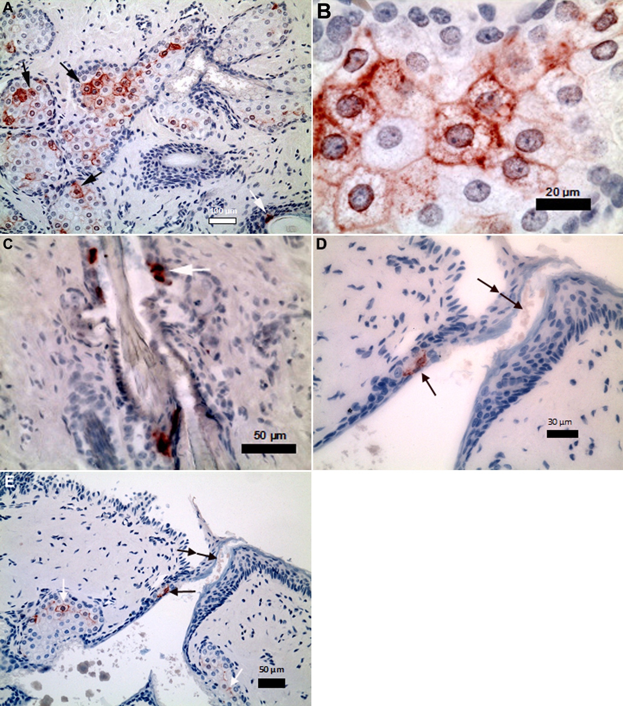
 Figure 1
of Cheriyan, Mol Vis 2011; 17:3055-3061.
Figure 1
of Cheriyan, Mol Vis 2011; 17:3055-3061.