Figure 3. Confocal images through the
mouse cornea 4 h after femtosecond laser keratotomy (FSL). A:
Healthy normal epithelial layer. B: Normal distribution
of sub-basal nerve fibers. C: Fragmentation and debris
of cellular matter in the region of the FSL injury. D:
Normal posterior corneal keratocytes. E: Healthy
hexagonal endothelial cells. All images were taken with the
Heidelberg HRT3 with the Rostock corneal module.
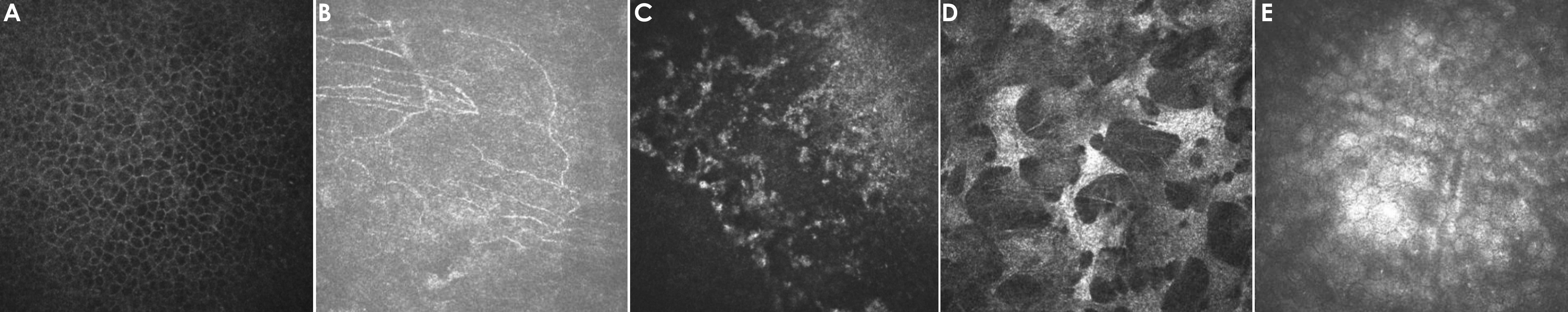
 Figure 3
of Angunawela, Mol Vis 2011; 17:3005-3012.
Figure 3
of Angunawela, Mol Vis 2011; 17:3005-3012.