Figure 1. Surgical positioning and post-operative images of mouse eye. A: Photo showing positioning of the mouse cornea against the treatment cone. The mouse eye was partially prolapsed and brought
into contact with the cone. The area of surface contact was visualized directly through the operating microscope. B: Color slit lamp image showing the mouse cornea 4 h after femtosecond laser (FSL) keratotomy . The circumferential outline
of the FSL incision is indicated by the arrow. The cornea is clear (note the clearly visible iris details) but with some corneal
edema present at this time point. A cataract is present behind the cornea and appears opaque (cyan arrow).
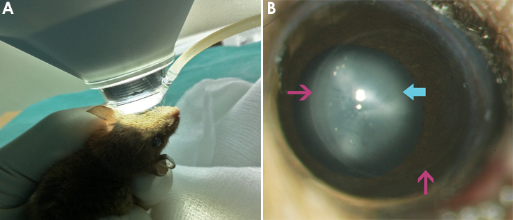
 Figure 1 of
Angunawela, Mol Vis 2011; 17:3005-3012.
Figure 1 of
Angunawela, Mol Vis 2011; 17:3005-3012.