Figure 1. Cloning of a novel FRMD7
isoform and tissue distribution of the two splice variants. A:
Components of the FERM domain and gene structure of hFRMD7-FL.
B and C: Sequence comparison between hFRMD7-FL/mFRMD7-FL
(B) and hFRMD7-S/mFRMD7-S (C)
showing the deletion of 45 bp in the 5′ end of exon 4 in the hFRMD7-S/mFRMD7-S.
D: Agarose gel electrophoresis of the RT–PCR products to
confirm their size and the identification of a single PCR
product. E: Expression levels of hFRMD7-FL and hFRMD7-S
transcripts in selected human fetal tissues. The primer sets
used in D and E: p1f/p1r, p2f/p2r and p3f/p3r.
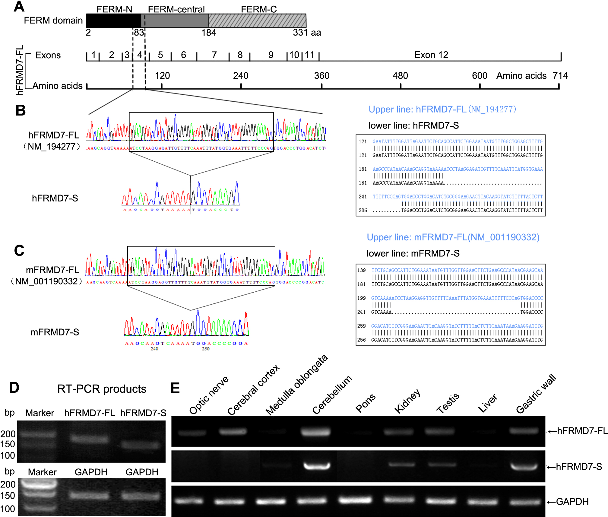
 Figure 1
of Li, Mol Vis 2011; 17:2986-2996.
Figure 1
of Li, Mol Vis 2011; 17:2986-2996.