Figure 9. Cy3-FluoSpheres® were injectedbefore, and after separation of the optic vesicle lumen from the brain ventricular system. These photographs
were taken 10 min after injection into the diencephalic ventricle. A: The older stage 31 (upper) embryo has a more advanced overall body shape, with fuller development of its tail bud (*) compared
to the stage 29 embryo beneath it. The arrows show the optic vesicles. B: Under fluorescence, although brain ventricle labeling is seen in both embryos, optic vesicle filling is seen only in the
stage 29 embryo (arrows).
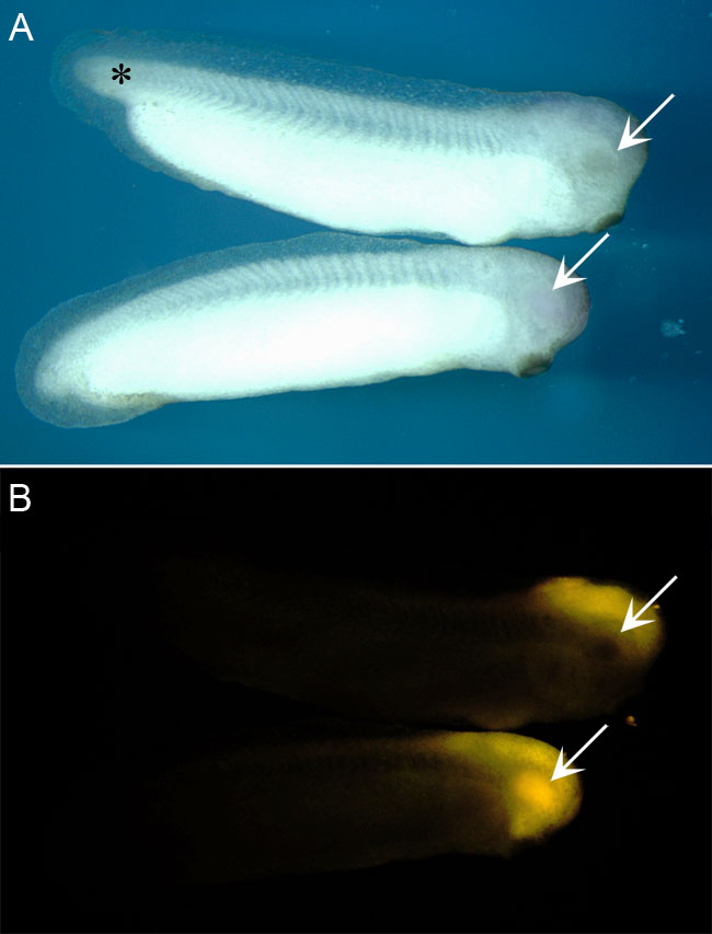
 Figure 9 of
Gonzalez-Fernandez, Mol Vis 2011; 17:2956-2969.
Figure 9 of
Gonzalez-Fernandez, Mol Vis 2011; 17:2956-2969.