Figure 7. Texas red microsphere were delivered to retinal pigment epithelium/retina interface. The contralateral optic vesicle was injected
with the microspheres at stage 26. A: At stage 28, the embryo was embedded in methacrylate polymer. At this stage, formation of the optic cup is complete and
melanin pigment is visible in the retinal pigment epithelium. B: The same section photographed under fluorescence shows the microspheres appearing as red dots (arrows) along the RPE/retina
interface.
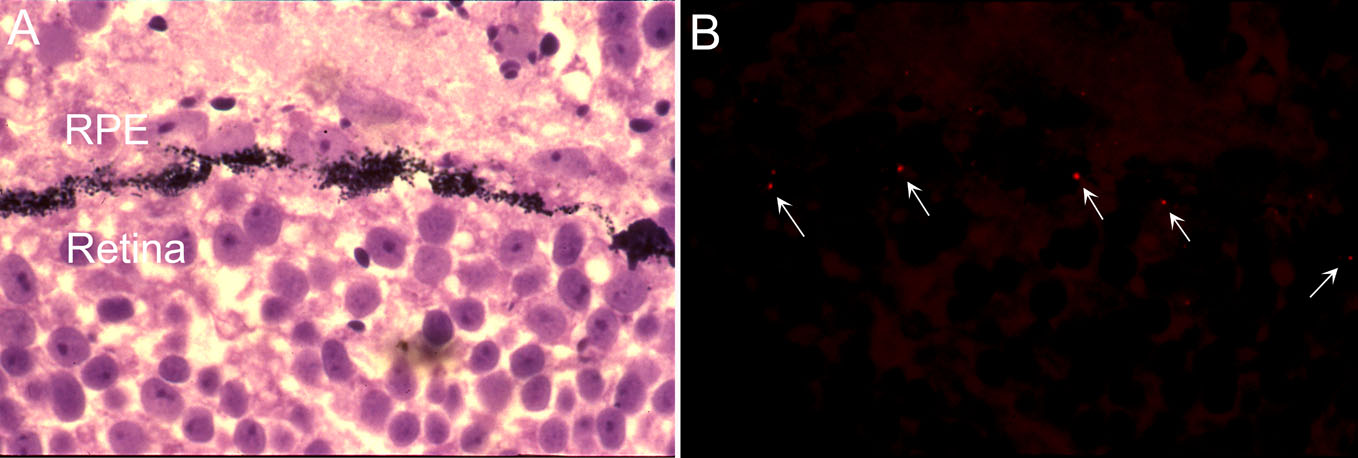
 Figure 7 of
Gonzalez-Fernandez, Mol Vis 2011; 17:2956-2969.
Figure 7 of
Gonzalez-Fernandez, Mol Vis 2011; 17:2956-2969.