Figure 6. Cy3 microspheres were injected into the diencephalic ventricle at stage 26. Panels A, B and C, D are dorsal and lateral views, respectively, of the same embryo showing the optic vesicles, which normally appear to bulge
laterally from the head (slanted arrows). Under fluorescence, the optic vesicles are filled with FluoSpheres® (panels B, D). The vertical arrow in panels C, D indicates the diencephalic ventricle which also contains the FluoSpheres®. The scale bar in the lower right equals 0.5 mm.
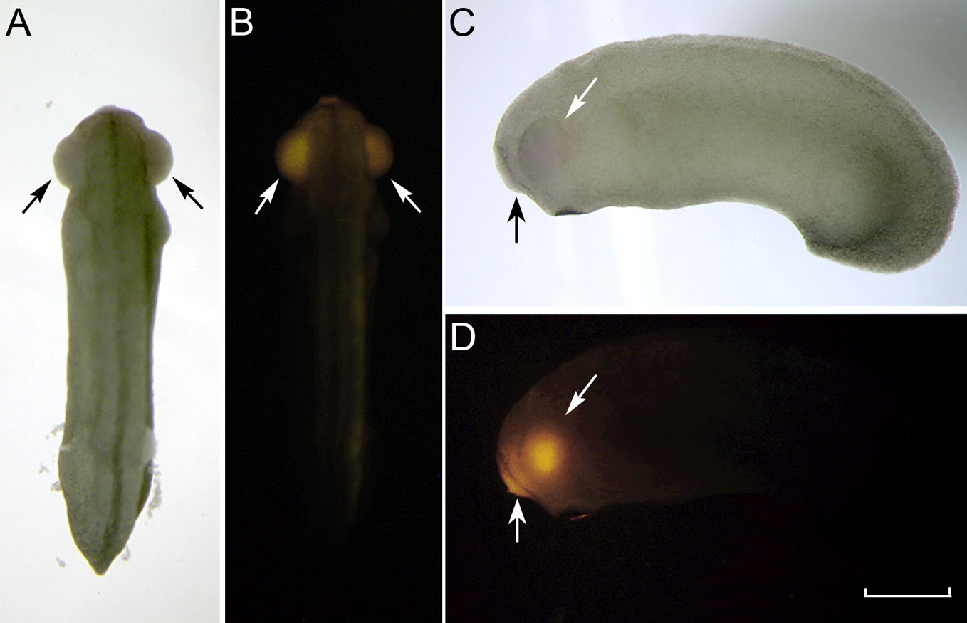
 Figure 6 of
Gonzalez-Fernandez, Mol Vis 2011; 17:2956-2969.
Figure 6 of
Gonzalez-Fernandez, Mol Vis 2011; 17:2956-2969.