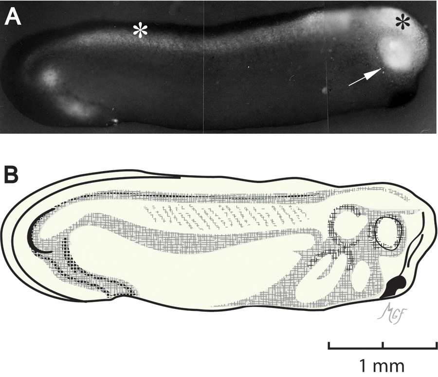
Figure 5. Optic vesicle fluorescein isothiocyanate–dextran injection. The injection was performed at stage 24, and photographed here
at stage 28.
A: Fluorescence shows filling of the optic vesicle (arrow), brain ventricle (black asterisk), and central canal (white asterisk).
B: This illustration was prepared to be consistent with stage 28 as in reference [
30].
 Figure 5 of
Gonzalez-Fernandez, Mol Vis 2011; 17:2956-2969.
Figure 5 of
Gonzalez-Fernandez, Mol Vis 2011; 17:2956-2969.