Figure 11. Diencephalic ventricular injection was used to deliver Cy3-FluoSpheres® to the retina of transgenic tadpoles expressing green fluorescent protein under the rod-opsin promoter. The brain ventricle
was injected through the anterior prosencephalon at embryonic stage 27, and development allowed to continue for 48 h to stage
41. A: In vivo photograph showing the normal external appearance of the eye and pupil (arrow). B: Cy3-FluoSphere® labeling of the retina can be viewed through the pupil. C: Retinal expression of green fluorescent protein (GFP) is apparent through the pupil. D: Cryosection (transverse plane) through the eye at the level of the optic nerve (arrowhead) shows that the Cy3-FluoSpheres® have diffusely labeled the diencephalon (asterisk), and are found extending through the optic nerve to the subretinal region.
E: This higher magnification of boxed area in panel D shows GCL, ganglion cell layer; inner nuclear layer, ONL, outer nuclear layer; OS, outer segments; RPE, retinal pigmented
epithelium; Arrowheads, rod photoreceptors expressing GFP.
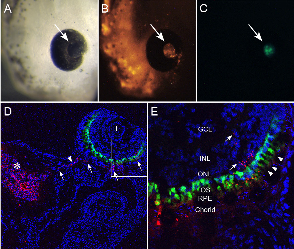
 Figure 11 of
Gonzalez-Fernandez, Mol Vis 2011; 17:2956-2969.
Figure 11 of
Gonzalez-Fernandez, Mol Vis 2011; 17:2956-2969.