Figure 3. RGCs transfected with YFP
using the modified capillary gun remain morphologically intact
for 1 week ex vivo. A: Transfection with the modified
capillary gun preserves RGC axons that extend from somata to the
optic nerve head. B: In contrast, transfection with a
conventional entrainment biolistic device produces many axons
that lose continuity mid-explant (arrows). C-E:
RGC axons remain healthy 2 (C), 4 (D), and 6 (E)
days after transfection with the modified capillary gun. F-H: In
contrast, those from explants transfected with the entrainment
device are somewhat healthy on day 2 (F), but show overt
degeneration by 4 (G) to 6 (H) days after
transfection. I: Quantification of these results reveals
that transfection with the entrainment device causes
significantly greater degeneration than does transfection with
the modified capillary gun. *p<0.05 by ANOVA. J:
Smi-31 staining of explants transfected with the modified
capillary gun reveals organized, bundled axons. K: In
contrast, those in explants transfected with a conventional
entrainment device show a disorganized axonal pattern. Scale bar
represents 100 μm in A and B and 50 μm in C-K.
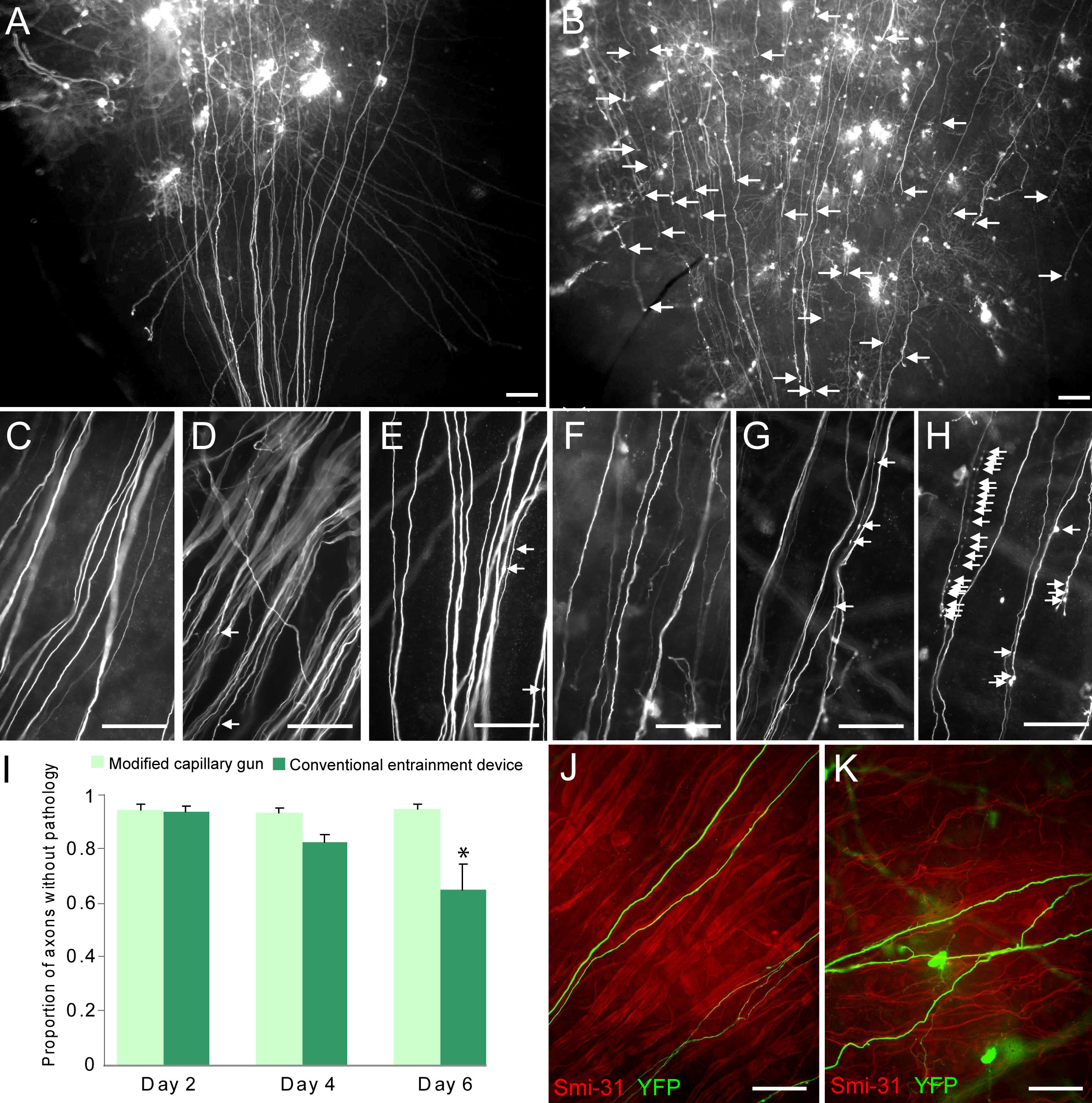
 Figure 3
of Christianson, Mol Vis 2011; 17:2947-2955.
Figure 3
of Christianson, Mol Vis 2011; 17:2947-2955.