Figure 2. Transfection with the
modified capillary gun labels retinal ganglion cells (RGCs) to
their distal tips. A, B: Transfection of a
retinal explant with YFP permits visualization of numerous RGCs
and their axons, which course toward the optic nerve head. C-F:
By 48 h after transfection, RGC somata and dendrites are fully
labeled with fluorescent protein and show no evidence of blebs
or degeneration after transfection with the modified capillary
gun. G-I: The micro-targeting capacity of the gun
permits clear visualization of YFP-filled RGC axons, which are
smooth, straight, and devoid of varicosities in the region near
the optic nerve head. Scale bar represents 100 μm in A
and B and 50 μm in C-I.
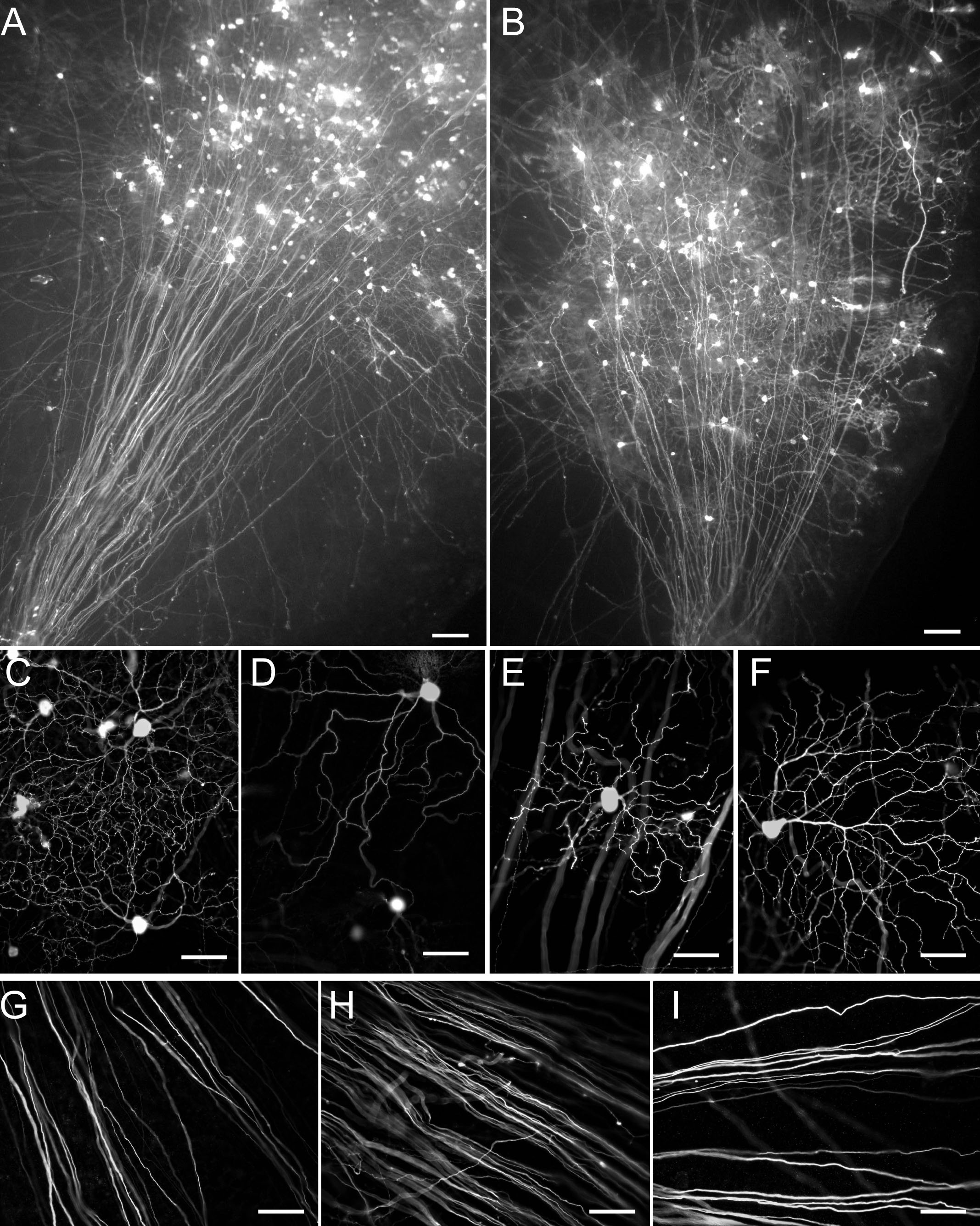
 Figure 2
of Christianson, Mol Vis 2011; 17:2947-2955.
Figure 2
of Christianson, Mol Vis 2011; 17:2947-2955.