Figure 5. Ablation of cell division cycle 42 homolog (S.cerevisiae; CDC42) does not affect progression of photoreceptor degeneration.
A: Retinal morphologies of Cdc42 conditional knockdown (cKD) and control mice before (dark control) or at 24 h and 10 days after light exposure are shown.
B: Photoreceptor apoptosis was quantified in Cdc42 knockdown (black bars) and wild-type controls (white bars) 36 h after light exposure. Shown are results from n=4 retinas
per time point and genotype. Statistical analysis showed no significant difference in apoptosis between wild-type and knockdown
mice (Student t-test). C: Retinal morphologies of Cdc42flox/flox;opsin-Cre; VPP (cKD VPP) and Cdc42flox/flox;VPP (control VPP) mice were examined at 4 weeks (weeks), 12 weeks and 20 weeks of age. Shown are representative sections
of n=3. D: Thickness of the outer nuclear layer (ONL) of Cdc42flox/flox;opsin-cre;VPP (cKD) and Cdc42flox/flox;VPP (ctrl) mice at 4, 12, and 20 weeks of age is shown in a spider diagram. Measurements from morphological sections of 3
mice per genotype and age are shown. Retinal thickness of wild-type and knockdown mice were comparable at all time points
and locations (ANOVA [ANOVA] followed by bonferroni post-hoc test). OPL: outer plexiform layer, INL: inner nuclear layer,
IPL; inner plexiform layer, GCL: ganglion cell layer, IS: photoreceptor inner segment, OS: photoreceptor outer segment. Scale
bars: 50 mm.
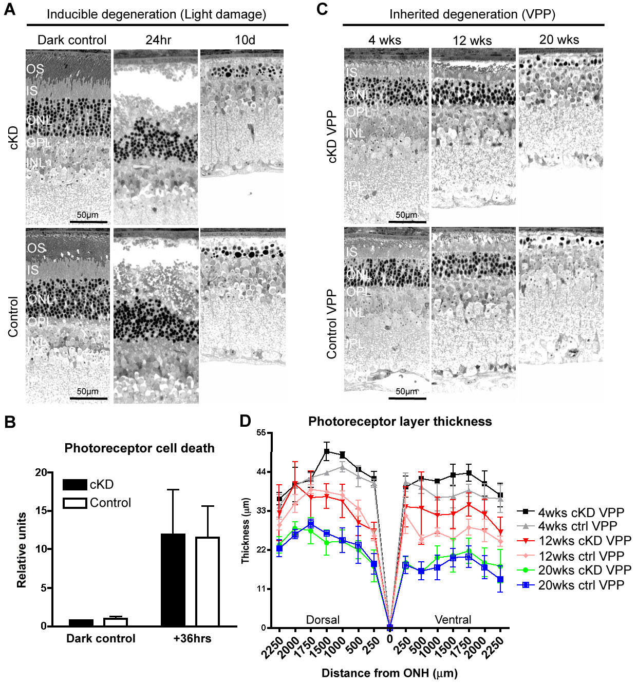
 Figure 5 of
Heynen, Mol Vis 2011; 17:2934-2946.
Figure 5 of
Heynen, Mol Vis 2011; 17:2934-2946.