Figure 3. Cell division cycle 42
homolog (S.cerevisiae; Cdc42) conditional knockdown was
achieved specifically in rod photoreceptor cells. A: PCR
products after amplification of the floxed region of Cdc42
from retinal genomic DNA of Cdc42 conditional knockdown
(cKD) and control mice at 12-weeks of age were separated and
visualized by DNA agarose electrophoresis. Amplification of the
floxed sequence results in a 1.5 kb fragment and of the excised
sequence in a 0.9 kb product. B: Relative expression of
Cdc42 in the outer nuclear layer (ONL; white box), the
inner nuclear layer (INL; black box), and the ganglion cell
layer (GCL; gray box) of Cdc42 knockdown and control
mice was analyzed by real-time PCR after laser capture
microdissection. Shown are mean values±SD of 3 independent mice.
Expression of Cdc42 in each retinal layer of control
mice was set to ‘1’. C: Retinal sections from Cdc42
knockdown and control mice 14 h after light damage were
immunofluorescently stained for CDC42. Images are
representatives of 3 independent mice per genotype. Red: CDC42.
Blue: nuclei (4’,6 diamidino-2-phenylindole [DAPI] staining).
Scale: 20 μm. OS: photoreceptor outer segments. IS:
photoreceptor inner segments.
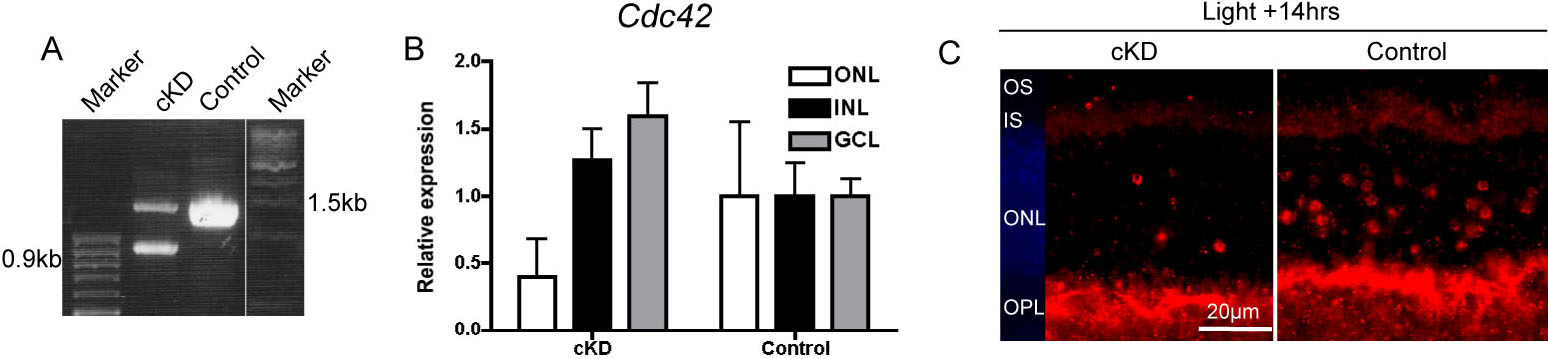
 Figure 3
of Heynen, Mol Vis 2011; 17:2934-2946.
Figure 3
of Heynen, Mol Vis 2011; 17:2934-2946.