Figure 1. Cell division cycle 42
homolog (S.cerevisiae; CDC42) localizes to the perinuclear
region of photoreceptors during retinal degeneration. A:
CDC42 was immunofluorescently labeled in retinal sections of
dark control mice and in mice at 14 h and 24 h after light
exposure. The first panel shows the control of a light exposed
retina (at 14 h after exposure) stained with the secondary
antibody alone. Shown are representative stainings of n=3 mice.
B: The outer nuclear layer (ONL) of CDC42 stained retinal
sections of mice at 14 h after light exposure, were analyzed by
confocal microscopy. The boxed area is shown at higher
magnification in the right panel. C: Retinal sections of
retinal degeneration (rd)1, rd10, autosomal
dominant retinitis pigmentosa (VPP) and wild-type age matched
controls at indicated post-natal days were immunofluorescently
stained for CDC42. D: Relative gene expression of Cdc42
was analyzed in retinas of dark controls (DC) and in retinas at
14 h and 24 h after light exposure. Shown are mean values±SD of
3 independent mice. E: Retinal layers were isolated by
laser capture microdissection and examined for Cdc42
expression. Gnat1 (ONL), Chx10 (inner nuclear
layer; INL), Opn4 (ganglion cell layer; GCL) served as
controls to assess purity of isolated layers and Actb was
amplified as a loading control. Blue: nuclei (4’,6
diamidino-2-phenylindole [DAPI] staining). Red: CDC42. Scale
bars 50 μm. RPE: retinal pigment epithelium, IPL: inner
plexiform layer, IS: photoreceptor inner segment, PND:
post-natal day.
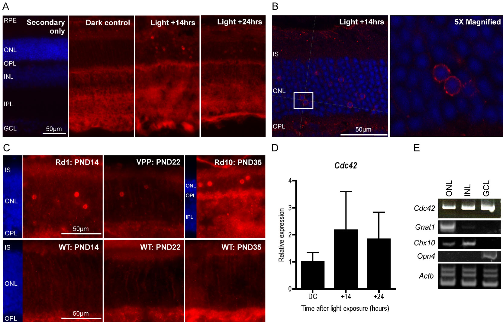
 Figure 1
of Heynen, Mol Vis 2011; 17:2934-2946.
Figure 1
of Heynen, Mol Vis 2011; 17:2934-2946.