Figure 8. Viability Staining of LECs cultivated over PCL nanofibers. A: LECs depicted high ratio of viable cells as demonstrated by positive green staining. Phase contrast micrograph shows that
LECs are migrating from the periphery of viable limbal explant (white arrowhead; 40× magnification). B: LECs cultivated on electrospun nanofibers shows confluent viable cell sheet at 100× magnification. C: Confocal microscopy depicted LESCs infiltrated the nanofibers and formed viable 3D corneal epithelium, positive viability
staining (green) nanofibers (gray).
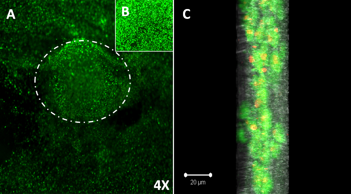
 Figure 8 of
Sharma, Mol Vis 2011; 17:2898-2910.
Figure 8 of
Sharma, Mol Vis 2011; 17:2898-2910.