Figure 7. Culture of limbal
epithelial cells cultivated on electrospun PCL nanofibers by
explant method. A: Confluent limbal epithelial cell
sheet grown on PCL nanofibers with round and ovoid morphology
examined using phase contrast microscopy at day 14 (100×
magnification). B: SEM image, LECs’ growth initiated
from the edge of the explant (black arrowhead). C: SEM
image showing that epithelial cells are closely attached to each
other with tightly opposed cell junctions, and D: apical
surface showing numerous short microvilli.
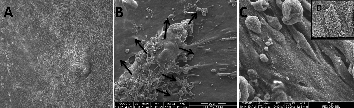
 Figure 7
of Sharma, Mol Vis 2011; 17:2898-2910.
Figure 7
of Sharma, Mol Vis 2011; 17:2898-2910.