Figure 6. SEM of HCE-T cells seeded
over nanofibers to evaluate the attachment ability at different
time intervals. A: SEM after 6 h post seeding shows
almost no cell attachment. B: After 1st day, cells
attached to the polymer surface, become large and flat in
morphology. C: After 3rd day, SEM revealed confluent
monolayer formation over nanofibers with good cell spreading. D:
Micrograph on the 5th day depicted good cell attachment and
spreading on the nanofibers surface. E: Micrograph on
day 7 illustrated confluent epithelial layer over nanofibers
surface similar as day five.
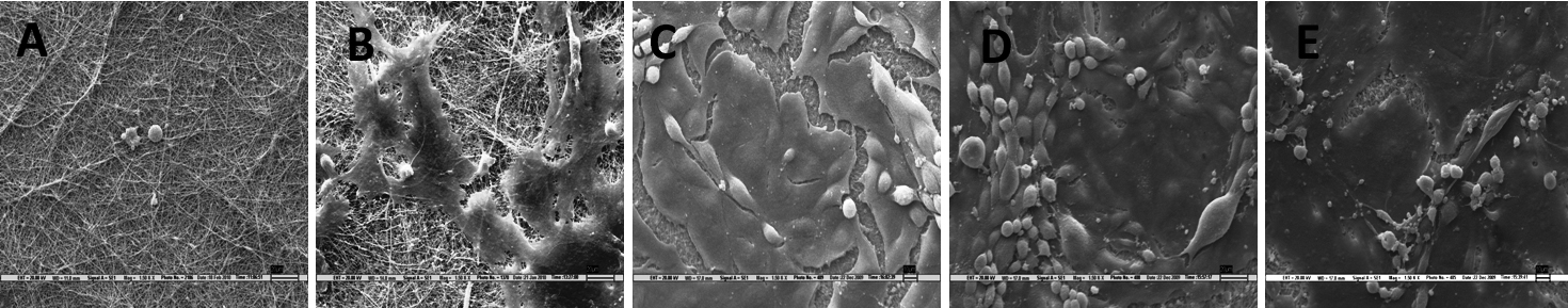
 Figure 6
of Sharma, Mol Vis 2011; 17:2898-2910.
Figure 6
of Sharma, Mol Vis 2011; 17:2898-2910.