Figure 5. Biocompatibility assessment of electrospun PCL nanofibers: A: Phase contrast pictures shows migration of HCE-T cells over nanofibers (black stars line). B: Epithelial cell sheet demonstrates high viability ratio of HCE-T cells on nanofibers by their positive green staining. Cells
were observed at 200× magnification.
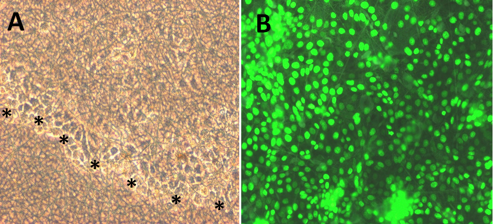
 Figure 5 of
Sharma, Mol Vis 2011; 17:2898-2910.
Figure 5 of
Sharma, Mol Vis 2011; 17:2898-2910.