Figure 5. Phosphorylation of Tyr14 in CAV-1 in NTM-5 and GTM-3 cells. A: NTM and GTM cell lysates were immunoprecipitated by polyclonal antirabbit Ab against CAV-1 and the precipitate was suspended
in a 2× loading buffer (ABCam Immunoprecipitation Protocol) and western blotted with monoclonal antimouse CAV-1 Ab. B: The same blot was stripped and reprobed with monoclonal antimouse pTyr14-CAV-1 antibody. C: Lanes shown in A and B were scanned using KODAK MI Software. The amount of non-phosphorylated CAV-1 is similar in NTM-5 and GTM-3 samples (A), while the amount of CAV-1 phosphorylated at Tyr14 (P-Tyr14-CAV-1) is 2.3 times lower in GTM-3 cells. Arrowhead indicates
heavy chain immunoglobulins. The values indicated represent mean±SEM from four independent experiments.
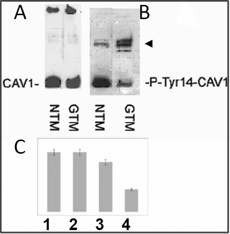
 Figure 5 of
Surgucheva, Mol Vis 2011; 17:2878-2888.
Figure 5 of
Surgucheva, Mol Vis 2011; 17:2878-2888.