Figure 3. Immunofluorescence localization of CAV-1 and CAV-2 in TM cells. CAV-1 (A, D, green). CAV-2 (B, E, red). NTM cells (A-C). GTM cells (D-F). Polyclonal rabbit anti-CAV-1 and monoclonal mouse anti-CAV-2 were used for immunostaining. C and F: merged; blue – DAPI staining. CAV-1 and CAV-2 immunoreactivity is identified as punctate staining with a predominant cytoplasmic
localization (arrows). In NTM cells CAV-1 was mainly found in the cytoplasm (A, arrows), while CAV-2 was present in cytoplasm, the perinuclear area as well as in the cell membranes (B, arrowheads). In GTM cells CAV-1 and CAV-2 are colocalized in dot-like structures (arrows, D-F).
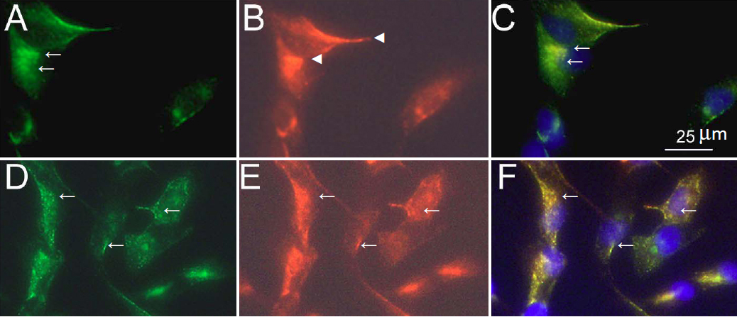
 Figure 3 of
Surgucheva, Mol Vis 2011; 17:2878-2888.
Figure 3 of
Surgucheva, Mol Vis 2011; 17:2878-2888.