Figure 7. Effect of photobleached
melanosomes on the sensitivity of individual ARPE-19 cells to H2O2-induced
toxicity compared to cells with a comparable content of control
particles (latex beads) in co-cultures. Live cell imaging of
dynamic changes in nuclear PI fluorescence over 18 h in ARPE-19
cells pre-loaded with latex beads or photobleached melanosomes
(pb mel) and treated with H2O2 delivered
as A: a pulse at 1500 μM or B: generated
enzymatically by the addition of GOx at 40 mU/ml. Numbers of
cells containing the different particle types selected for
analysis were as follows. A: beads, n=59 (triangles); pb
mel, n=63 (circles). B: beads, n=163 (triangles); pb
mel, n=74 (circles). Data are from representative experiments
and are the percent of the pre-selected cells in each particle
group surviving (no nuclear PI) with time. The shift to the left
of the curves illustrates the magnitude of the cytotoxic effect
of pb mel relative to beads. The curves for the two particle
types within each treatment protocol differ significantly
(GraphPad Prism 5 survival analysis, p<0.02).
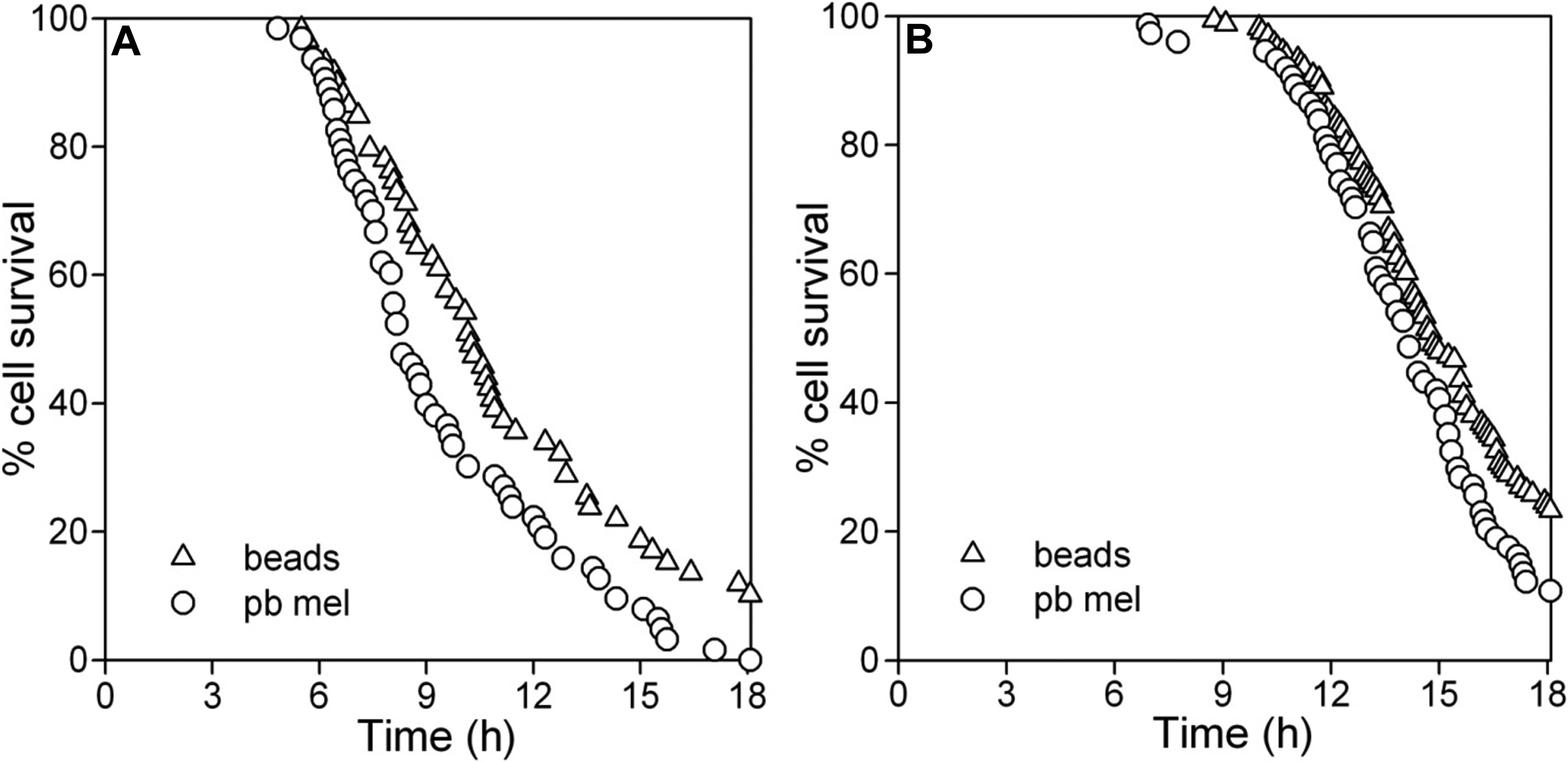
 Figure 7
of Burke, Mol Vis 2011; 17:2864-2877.
Figure 7
of Burke, Mol Vis 2011; 17:2864-2877.