Figure 4. Illustration of an imaging
assay using co-cultures of ARPE-19 cells pre-loaded by
phagocytosis with control latex beads or melanosomes. The same
microscope field is shown by A: bright-field microscopy,
and B, C: in merged bright-field and
fluorescence images. The fluorescence image overlay in B
shows latex bead autofluorescence (artificially colorized blue)
and the double overlay in C also shows PI fluorescence
(red), illustrating nuclear staining at 9 h and 20 min after
addition of 750 μM H2O2. Circled cells in
A show examples of cells pre-selected for analysis by
their particle content. In B, the same cells are circled
to illustrate discrimination of particle type in the selected
cells; one contains beads (arrow) and the other melanosomes
(double arrow). The arrow in C indicates a PI-positive
nucleus in the bead-containing cell after H2O2
treatment. Scale bar: 20 μm.
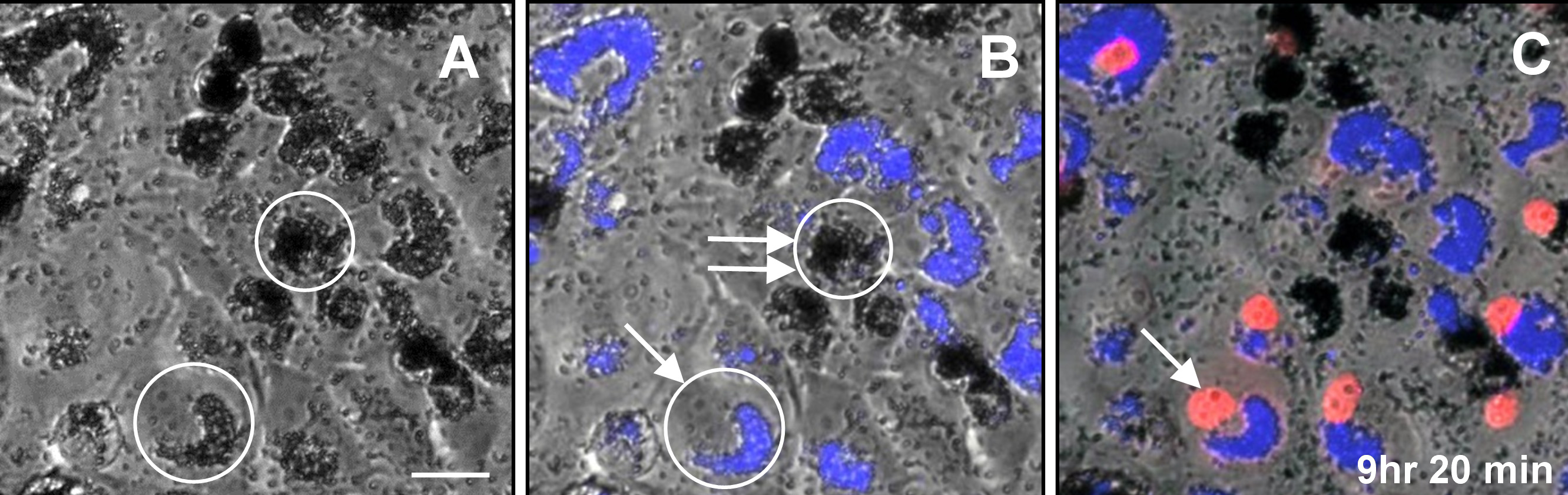
 Figure 4
of Burke, Mol Vis 2011; 17:2864-2877.
Figure 4
of Burke, Mol Vis 2011; 17:2864-2877.