Figure 4. Representative images for MUC5AC
staining in the conjunctiva. In the conjunctival fornix, more MUC5AC
positive cells in the control group (A) were observed than that
of the BAC-treated group (B), while none was recorded in the
bulbar conjunctiva near limbus (C). The average number of MUC5AC
positive cells was significantly decreased after BAC treatment (D).
**p<0.01.
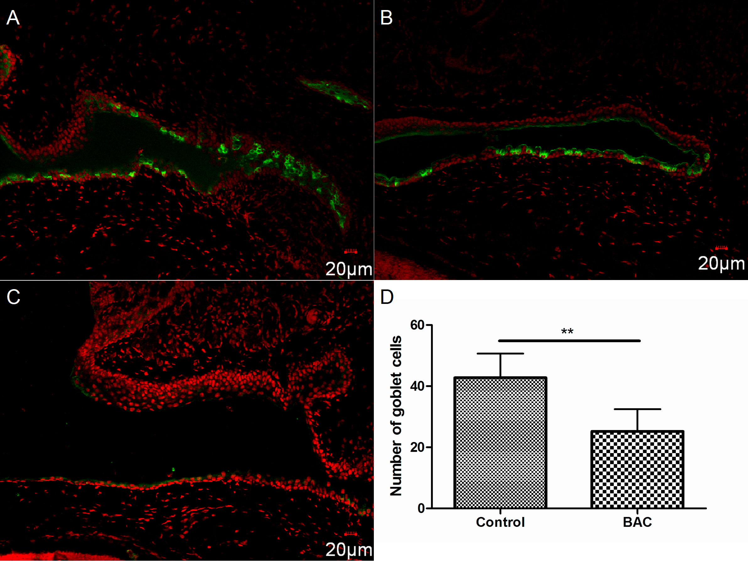
 Figure 4 of Lin, Mol Vis 2011; 17:257-264.