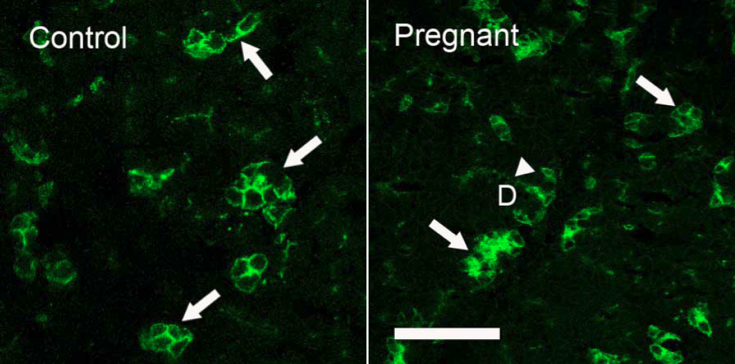
Figure 8. Immunofluorescence of AQP5-IR. Control: AQP5-IR was present in both basolateral and apical membranes of acinar cells, and
distributed among acini in a “mosaic” pattern, with some acini and/or acinar cells demonstrating much stronger AQP5-IR (arrows)
than the rest of acini/acinar cells. However, little AQP5-IR was detected in duct cells. These results were similar to our
previous reports [
5,
6]. Pregnant: as in control LG, AQP5-IR was also present in a “mosaic” pattern with a similar intensity and distribution pattern
(arrows). However, in contrast to control animals, ductal cells also exhibited a significant amount of AQP5-IR (arrowhead).
D=duct. Scale bar=50 μm.
 Figure 8 of
Ding, Mol Vis 2011; 17:2847-2855.
Figure 8 of
Ding, Mol Vis 2011; 17:2847-2855.