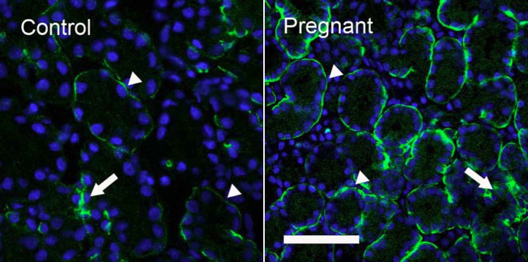
Figure 7. Immunofluorescence of AQP4-IR. Control: AQP4-IR was observed on the basolateral sides of acinar and duct cells, with duct
(arrow) showing much stronger AQP4-IR than acini (arrowheads), results in accordance with our previous reports [
5,
6]. Pregnant: Acini in pregnant rabbits showed substantially stronger basolateral staining (arrowheads) than control, whereas
AQP4-IR in ducts (arrow) appeared to be similar to that of control. In both images, DAPI was used to stain nuclei as bright
blue to demonstrate the morphologic profiles of acini and ducts. Scale bar=50 μm.
 Figure 7 of
Ding, Mol Vis 2011; 17:2847-2855.
Figure 7 of
Ding, Mol Vis 2011; 17:2847-2855.