Figure 5. Nuclear p38MAPK
localization is necessary for HCF migration. HCFs were seeded on
collagen in SSFM at 1×10⁵ cells/ml in a 24 well dish.
The next day cells were scratch-wounded and incubated in (A,
F, K) p38MAPK inhibitor, 10 μM SB202190, (B,
G, L) DMSO, (C, H, M)
TGFβ1 antibody, (D, I, N) Control IgG, or
(E, J, O) TGFβ RI (ALK5) inhibitor 10 μM
SB431542. After 4 h cells were fixed and immunostained for
p38MAPK (A-E) or SMAD 2/3 (F-J).
Arrows denote the nuclei of leading edge cells in which p38MAPK
and SMAD 2/3 were either localized or excluded (Bar=50 μm) or
after 18 h cells were imaged (K-O). Bar=200 μm.
DMSO is the control for addition of SB202190 or SB431542.
Quantification of all data are shown in the bar graphs below the
images. Left to right: Exclusion of p38MAPK from the nucleus in
leading edge cells, exclusion of SMAD 2/3 from the nucleus in
leading edge cells, cell migration into the wound. Nuclear
localization was counted using Image J software. Two non-biased
people scored cell migration, 0 (less migration) to 5 (most
migration). **p-value <0.01, ***p-value <0.001. NS=not
significant. Experiments were repeated at least three times with
similar results.
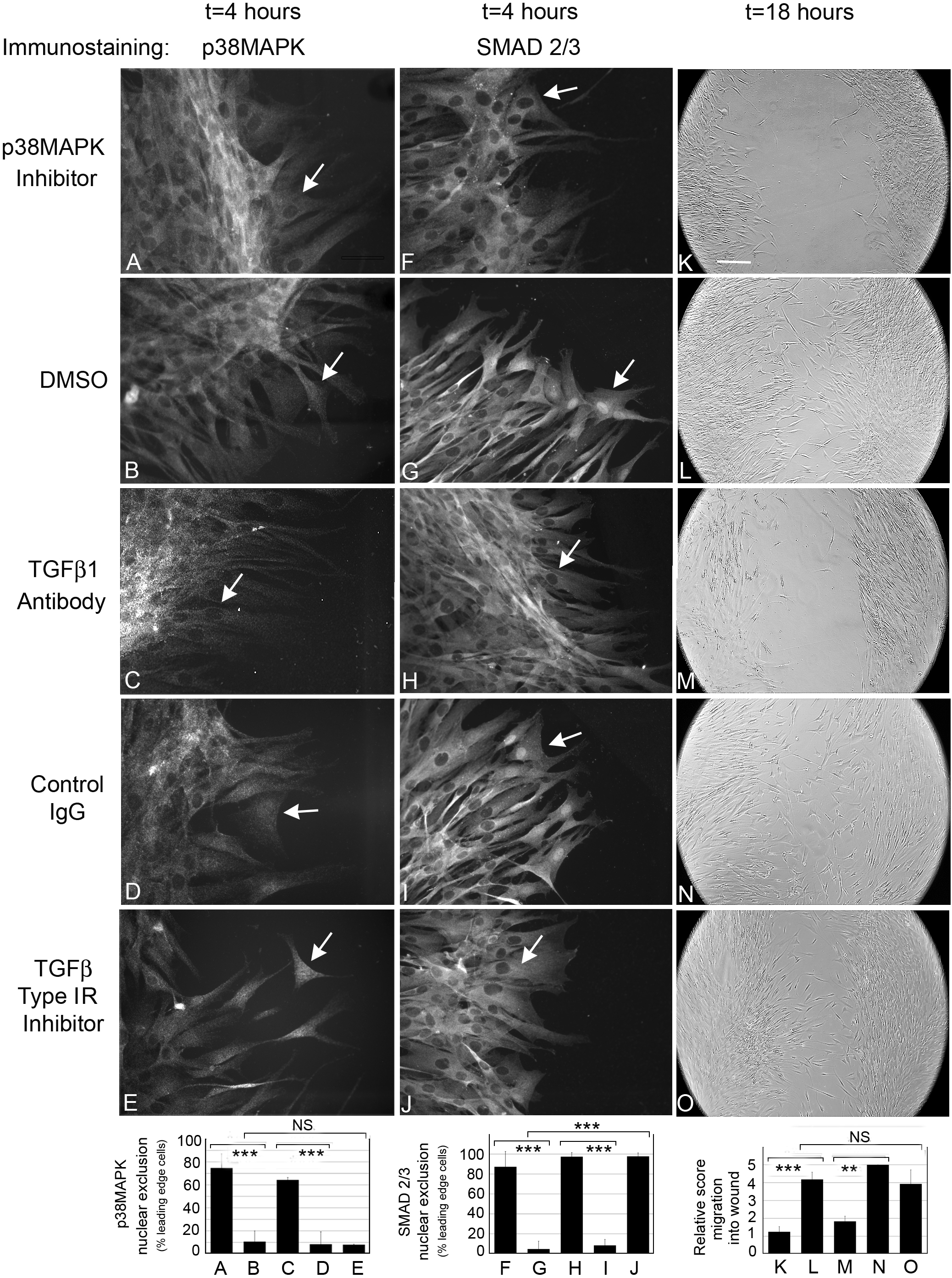
 Figure 5
of Wang, Mol Vis 2011; 17:2835-2846.
Figure 5
of Wang, Mol Vis 2011; 17:2835-2846.