Figure 4. Increasing TGFβ1
concentrations results in a loss of nuclear p38MAPK. HCFs were
seeded on collagen in SSFM at 1×10⁵ cells/ml in a 24
well dish. The next day cells were scratch-wounded and incubated
in (A, E) SSFM, (B, F) 0.01 ng/ml
TGFβ1, (C, G) 0.1 ng/ml TGFβ1, or (D, H)
1.0 ng/ml TGFβ. After 4 h cells were fixed and immunostained for
p38MAPK (A-D) or SMAD 2/3 (E-H).
Bar=50 μm. Arrows point to nuclei in which we detected either
the nuclear localization or nuclear exclusion of p38MAPK and
SMAD 2/3. The percent of cells in which p38MAPK was excluded
from nuclei of the leading edge cells is shown in (I).
The percent of cells with SMAD 2/3 concentrated in nuclei in the
leading edge cells is shown in (J). Image J software was
used for quantification. Each condition was compared to SSFM.
***p-value <0.001. Experiments were repeated at least three
times with similar results.
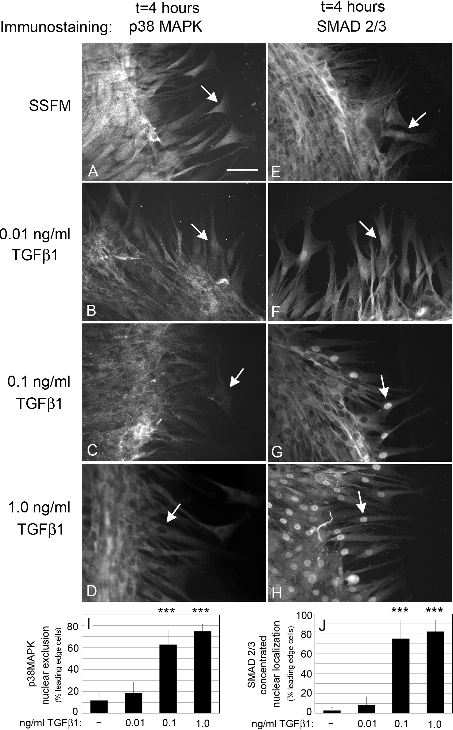
 Figure 4
of Wang, Mol Vis 2011; 17:2835-2846.
Figure 4
of Wang, Mol Vis 2011; 17:2835-2846.