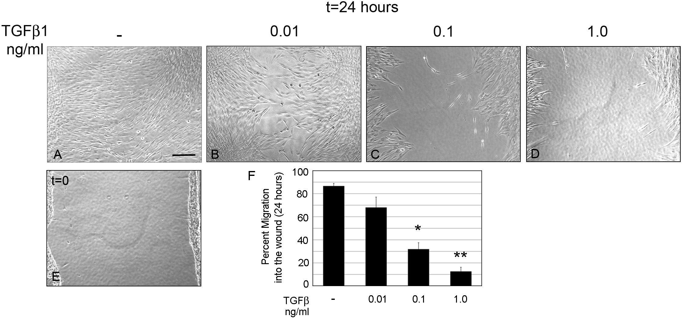
Figure 2. Increasing TGFβ1
concentrations reduces HCF migration. HCFs were seeded on
collagen in SSFM at 1×10
5 cells/ml in a 24 well dish.
The next day cells were scratch-wounded and incubated for 24 h
in (
A) SSFM, (
B) 0.01 ng/ml TGFβ1, (
C) 0.1
ng/ml TGFβ1, (
D) 1.0 ng/ml TGFβ1, or (
E) imaged at
time zero. Bar=200 μm.
F: Using
T-Scratch software,
percent cell migration into the wound margin at 24 h compared to
time zero was calculated. Each condition was compared to SSFM,
*p-value <0.05, **p-value <0.01. Experiments were repeated
at least three times with similar results.
 Figure 2
of Wang, Mol Vis 2011; 17:2835-2846.
Figure 2
of Wang, Mol Vis 2011; 17:2835-2846.