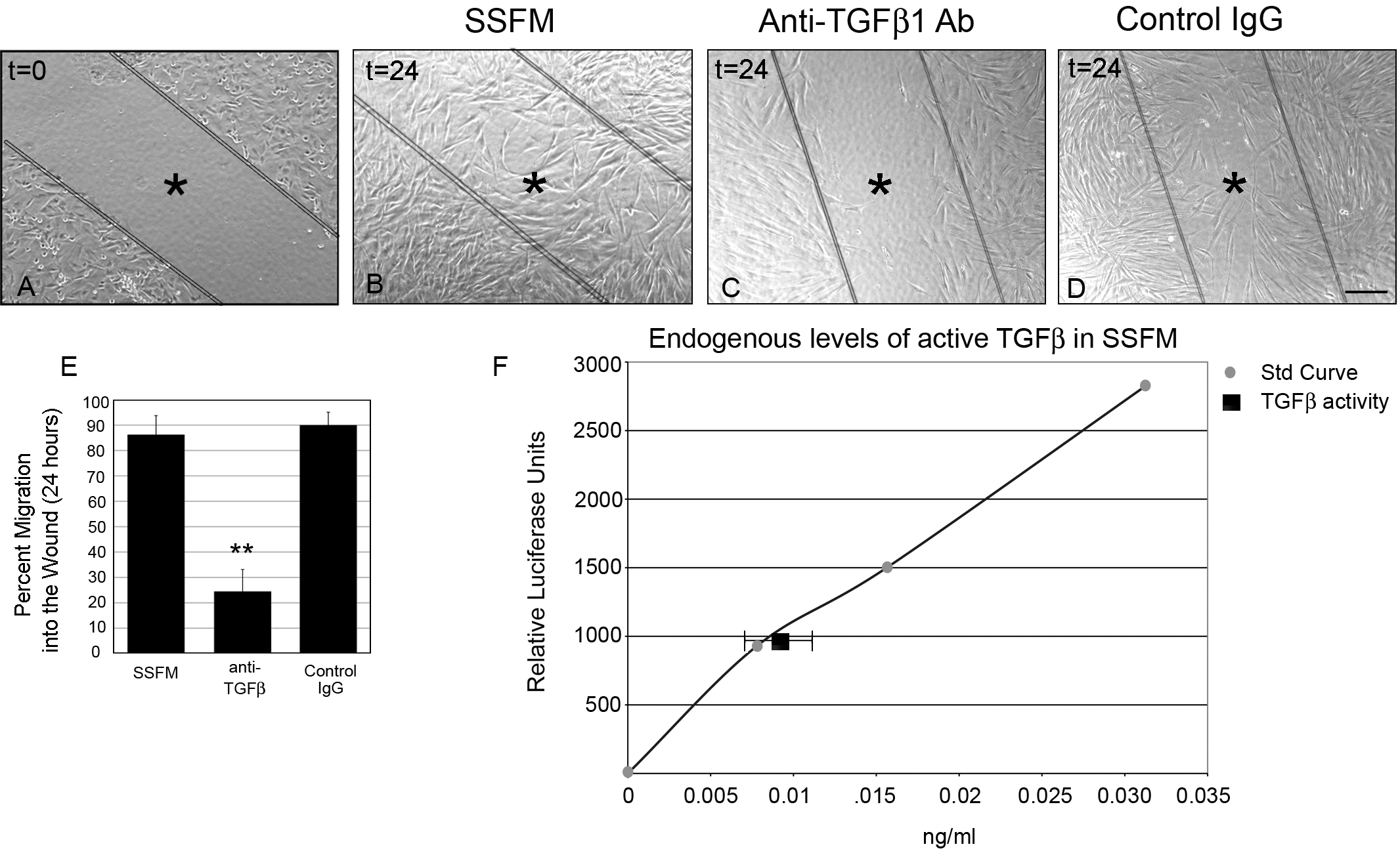
Figure 1. Endogenously secreted TGFβ1
promotes wound healing. HCFs were seeded on collagen in SSFM at
1×10
5 cells/ml in a 24 well dish. The next day cells
were scratch-wounded and imaged (
A) time zero, or
incubated for 24 h with either (
B) SSFM (
C) 2
μg/ml anti-TGFβ1 antibody or (
D) 2 μg/ml matched IgG
control. Bar=200 μm, *inside lines denoting wounded area.
E:
Using
T-Scratch
software, percent cell migration into the wound margin at 24 h
compared to time zero was calculated. Each condition was
compared to SSFM, **p-value <0.01.
F: To determine
endogenous levels of TGFβ, HCFs were co-cultured with TMLC,
which contain the PAI-1 promoter fused to the luciferase gene.
This assay demonstrated that HCFs have 0.01 ng/ml active TGFβ.
Experiments were repeated at least three times with similar
results.
 Figure 1
of Wang, Mol Vis 2011; 17:2835-2846.
Figure 1
of Wang, Mol Vis 2011; 17:2835-2846.