Figure 2. SDS–PAGE and western
immunoblot analysis of human lens membranes. Isolated cortical
and nuclear lens membranes were extracted with 7 M urea in Tris
buffer (CU – cortex urea; NU – nucleus urea) and with 0.1 M NaOH
(CA – cortex alkali; NA – nucleus alkali). Samples were analyzed
by SDS–PAGE (100 μg of membrane per lane) and with western
immunoblotting (20 μg of membrane per lane). Blots were probed
using anti-αA-crystallin, anti-αB-crystallin, anti-filensin and
anti-vimentin antibodies, as indicated. Molecular weight markers
are on the left of the figure.
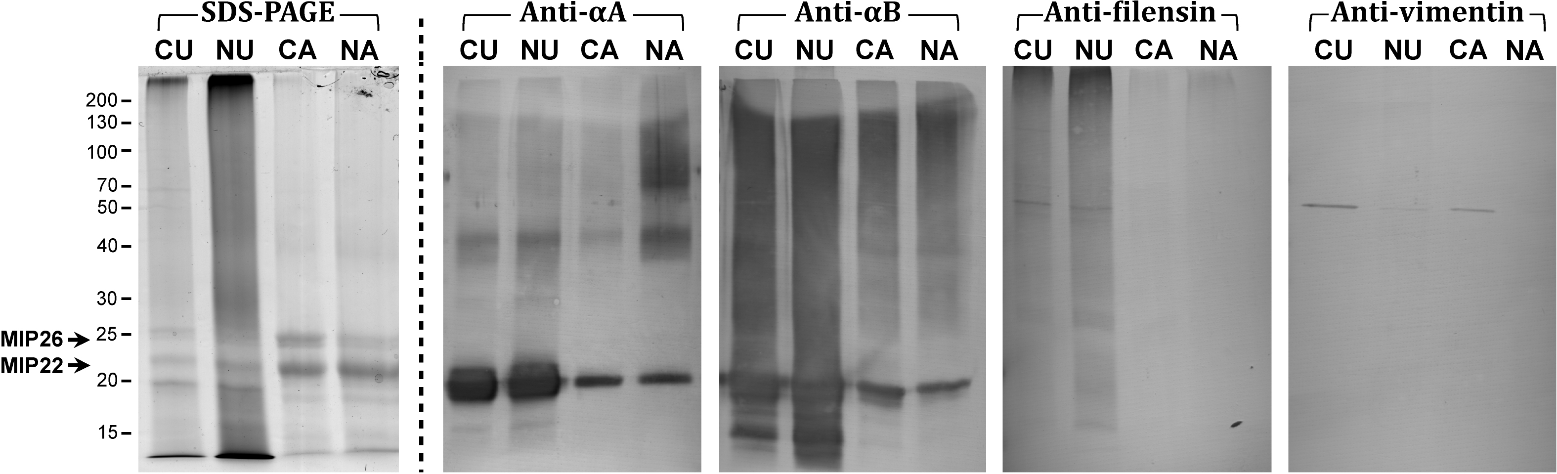
 Figure 2
of Su, Mol Vis 2011; 17:2798-2807.
Figure 2
of Su, Mol Vis 2011; 17:2798-2807.