Figure 1. SDS–PAGE and western
immunoblot analysis of bovine lens membranes. The
water-insoluble cell membrane pellets from bovine lens
homogenates washed with 7 M urea (BU) and with 0.1 M NaOH (BA).
Samples were analyzed by SDS–PAGE (100 μg of membrane per lane)
and western immunoblotting (20 μg of membrane per lane). Blots
were probed using anti-αA-crystallin, anti-αB-crystallin,
anti-filensin and anti-vimentin antibodies, as indicated.
Molecular weight markers are on the left of the figure.
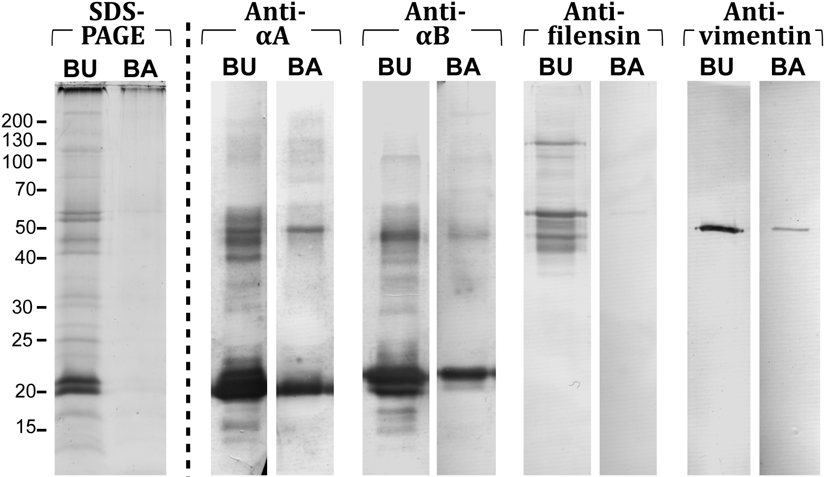
 Figure 1
of Su, Mol Vis 2011; 17:2798-2807.
Figure 1
of Su, Mol Vis 2011; 17:2798-2807.