Figure 2. Photomicrographs of rat
conjunctival epithelial cells (Cj-ECs) in culture. A, B:
Cj-ECs stained intensely for pan-keratin, a marker of
conjunctival epithelial cells. C, D:
Histochemical reactivity of primary culture of Cj-ECs to AB/PAS.
Goblet cells stained intensely with AB/PAS, indicating the
presence of both neutral (pink) and acidic (blue)
glycoconjugates associated with cells. Magnification: (A,
C), 100×; (B, D), 400×.
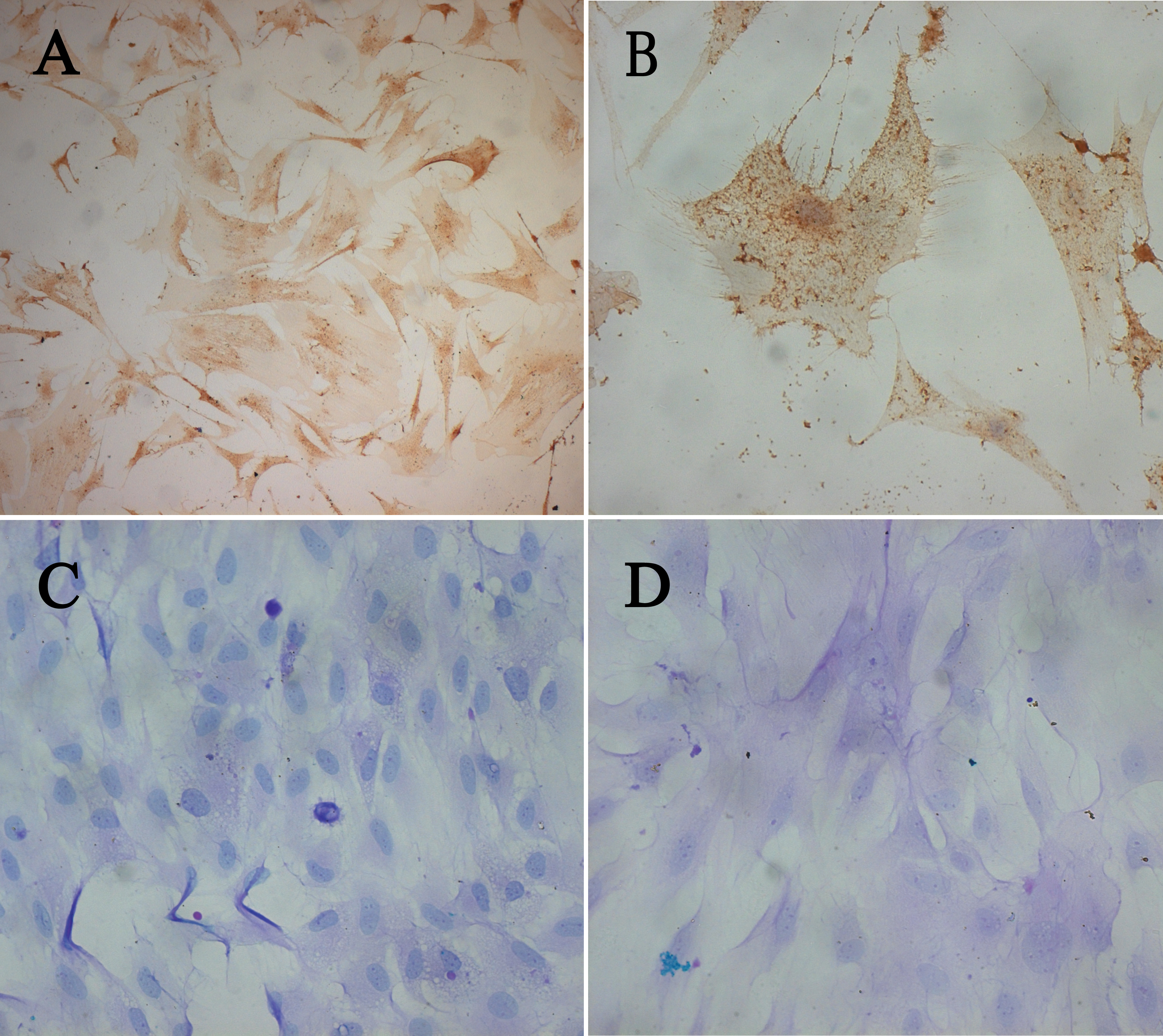
 Figure 2
of Ma, Mol Vis 2011; 17:2789-2797.
Figure 2
of Ma, Mol Vis 2011; 17:2789-2797.