Figure 1 of
Ma, Mol Vis 2011; 17:2789-2797.
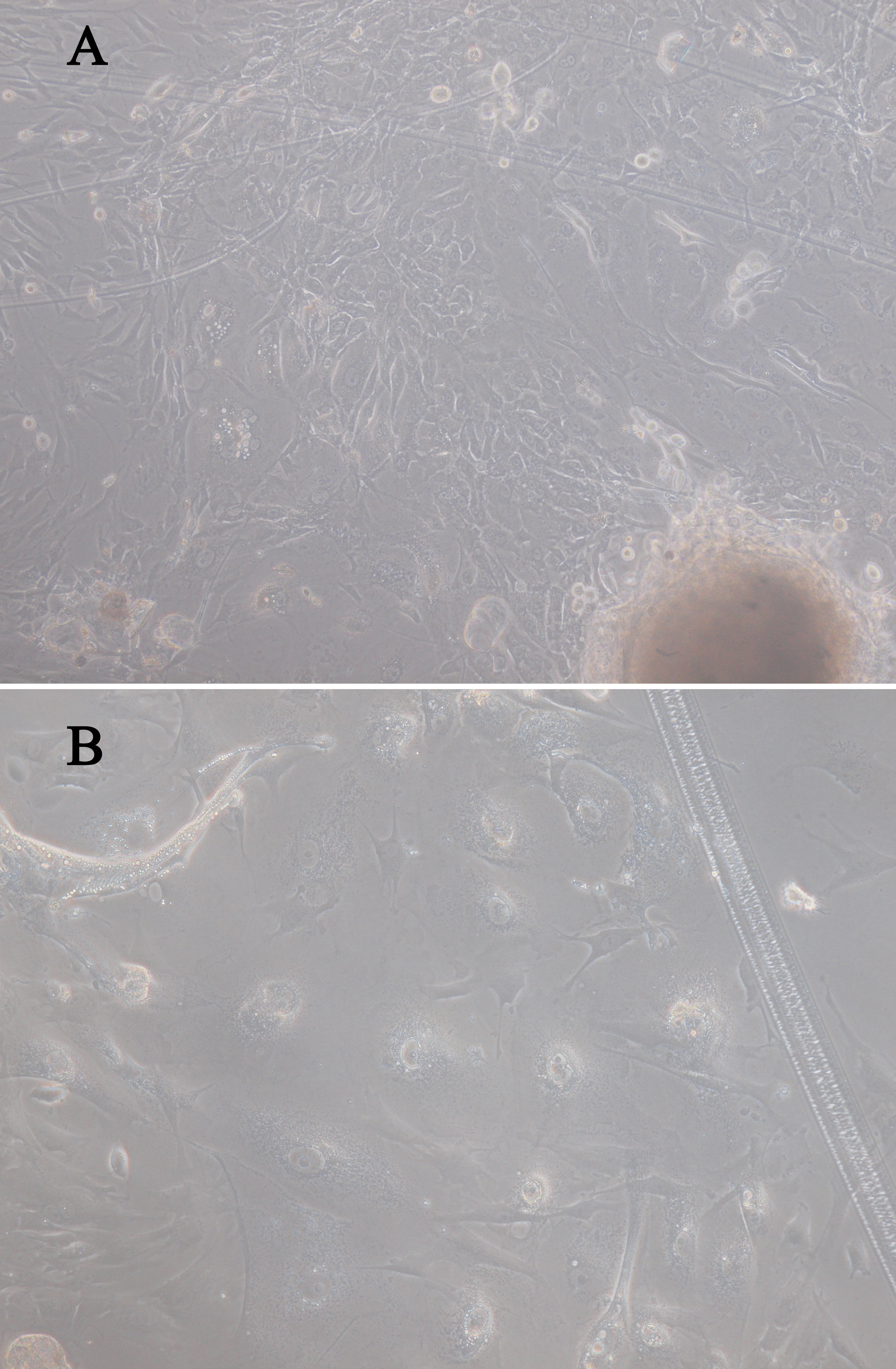
Figure 1.
Phase contrast micrographs illustrating the morphology of rat conjunctival explants grown in vitro. Magnification: (
A
) 100×, (
B
) 400×.
 Figure 1
of Ma, Mol Vis 2011; 17:2789-2797. Figure 1
of Ma, Mol Vis 2011; 17:2789-2797.
Figure 1
of Ma, Mol Vis 2011; 17:2789-2797. Figure 1
of Ma, Mol Vis 2011; 17:2789-2797.