Figure 10. Skp2 siRNA inhibited the proliferation of PFC and NFC in vivo. Hematoxylin and eosin staining was performed to examine the histological
changes 14 days after transfection. Obvious PFC and NFC proliferation was detected in pSuppressor vehicle group or without
transfection (A, D, B, E). There was little PFC and NFC proliferation in Skp2 siRNA transfection group (C, F). Scale bar is equal to 20 μm.
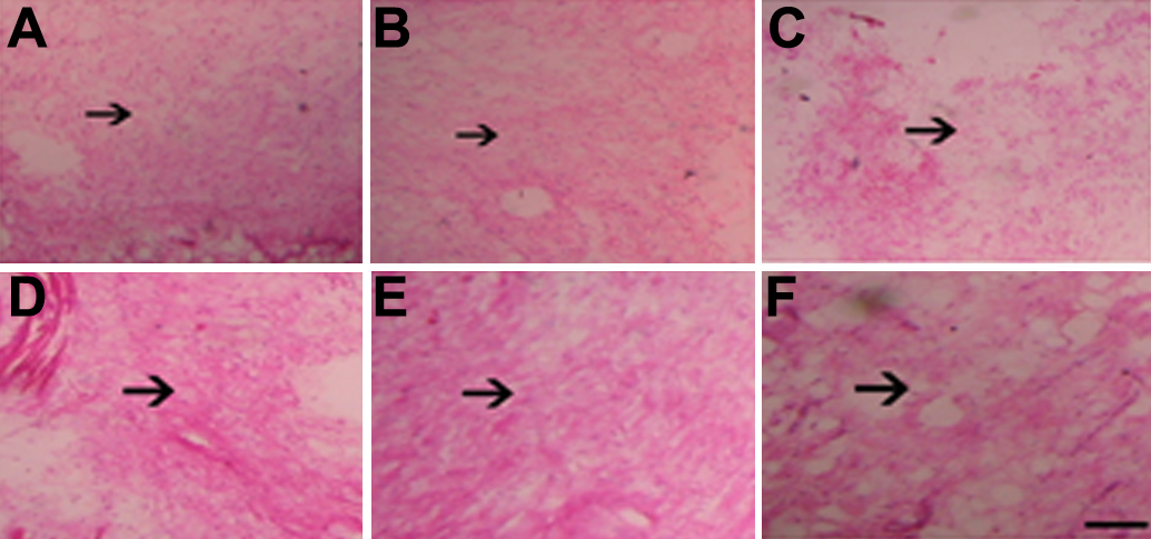
 Figure 10 of
Su, Mol Vis 2011; 17:247-256.
Figure 10 of
Su, Mol Vis 2011; 17:247-256.