Figure 6. Detection of melanopsin and
blue opsin in Moorfields/Institute of Ophthalmology-Müller 1
(MIO-M1) cells and human retinas by western blot analysis. A:
Melanopsin (Me; 65 kDa) was found in the cytosolic
(Cyt) and membrane (Mem) fractions of MIO-M1 cells. As the
control, the presence of melanopsin was detected in neural
retinal tissues from two postmortem donors (1, 2). The blots
were stained with (a) and without (b) the first antibody. B:
Presence of blue opsin (BO; 40 and 42 kDa) in the
cytosolic (Cyt) and membrane (Mem) fractions of MIO-M1 cells and
in two neural retinas (R1, R2), respectively (a). The isotype
control (b) was made with purified rabbit IgG.
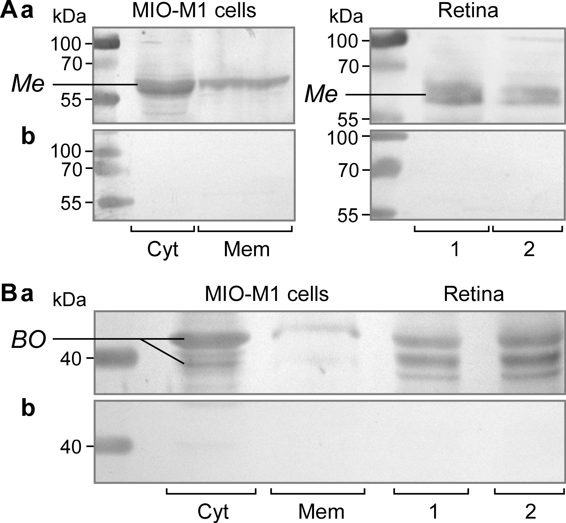
 Figure 6
of Hollborn, Mol Vis 2011; 17:2738-2750.
Figure 6
of Hollborn, Mol Vis 2011; 17:2738-2750.