Figure 5. Immunolabeling of opsins in
Moorfields/Institute of Ophthalmology-Müller 1 (MIO-M1) cells. A:
Labeling of blue opsin and melanopsin, respectively, in MIO-M1
cells. The cells were counterstained against glutamine
synthetase (GS) and GFAP, respectively. Double labeling yielded
a yellow-orange merge signal. B: Labeling of rhodopsin
and red-green opsin, respectively, in MIO-M1 cells. The cells
were counterstained against GFAP. C: Control culture of
MIO-M1 cells that was stained with secondary antibodies (goat
antirabbit IgG and goat antimouse IgG) and without primary
antibodies. No nonspecific labeling was observed either,
following incubation with goat antirat IgG (not shown). D:
Immunostaining of HEK-293 cells against melanopsin, blue opsin,
and red green opsin, respectively. The cells were counterstained
against glutamine synthetase (GS). Cell nuclei were labeled with
the DNA dye Hoechst 33258 (blue). Bars are 20 µm.
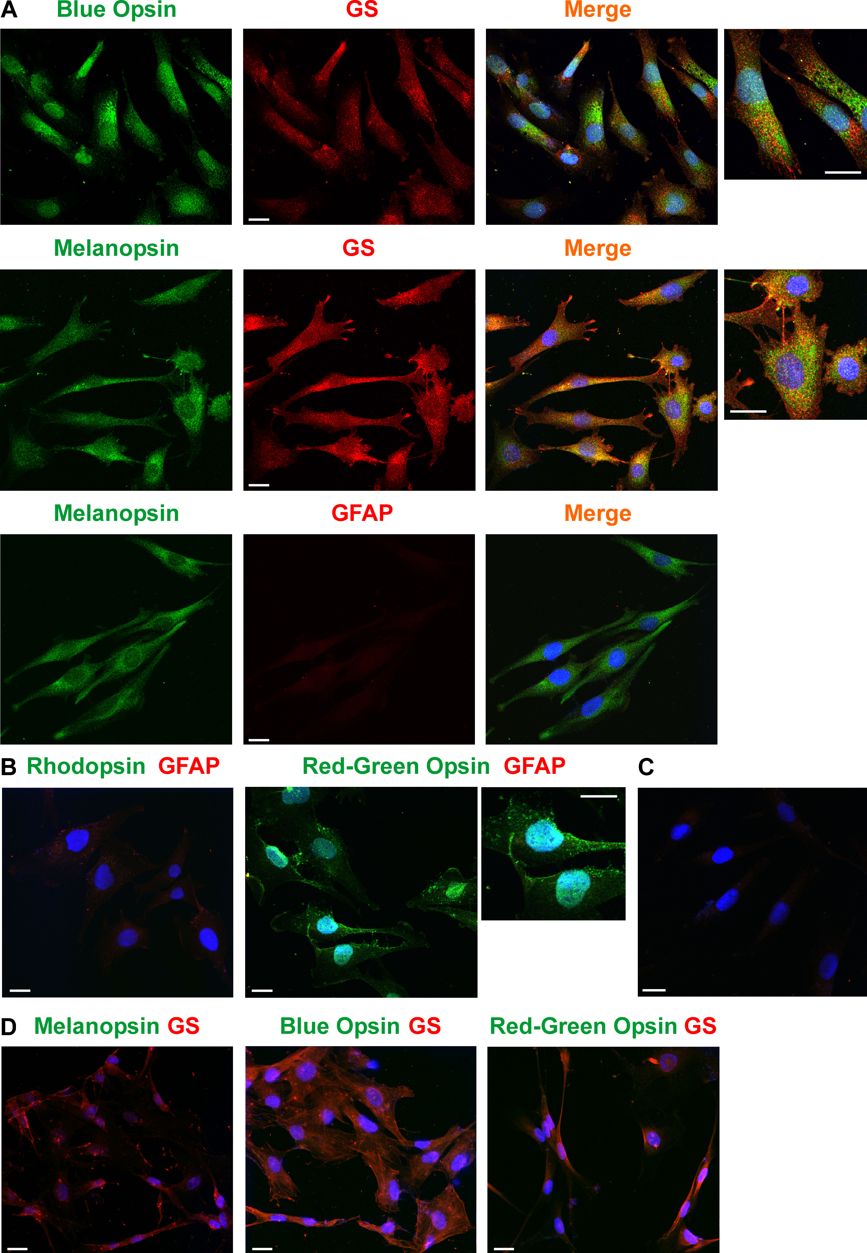
 Figure 5
of Hollborn, Mol Vis 2011; 17:2738-2750.
Figure 5
of Hollborn, Mol Vis 2011; 17:2738-2750.