Figure 3. mRNA expression of opsins
in Moorfields/Institute of Ophthalmology-Müller 1 (MIO-M1) cells
and human retinas. A: The expression of the following
genes was determined with real-time PCR in MIO-M1 cells and (B)
in neural retinal tissues from post-mortem donors. Long- and
midwave-sensitive cone opsin (OPN1LW+MW),
shortwave-sensitive cone opsin (OPN1SW), rhodopsin (OPN2),
panopsin (OPN3), melanopsin (OPN4), neuropsin (OPN5),
retinal G protein-coupled receptor (RGR), and peropsin (RRH).
In addition, the detection of hypoxanthine
phosphoribosyl-transferase (HPRT) transcripts is shown.
The data from two independent experiments (1, 2) are shown. The
negative control (-) was done by adding double-distilled water
instead of cDNA. C: A plasmid (pBSII SK+OPN4)
containing the open reading frame of melanopsin (OPN4; 1)
was used as positive control for the PCR experiments. D:
MIO-M1 cells that were cultured for 18 h in the absence (1) and
presence (2), respectively, of fetal bovine serum (10%)
contained transcripts for OPN4. E: Relative
expression level of opsins in MIO-M1 cells and postmortem neural
retinal tissues. The data were determined by real-time-PCR. The
bars represent the cycle numbers required to detect the
transcripts (relative to the cycle number for the detection of HPRT
mRNA). The data are the means±standard error of the mean (SEM)
of seven independent experiments. *P value (p)<0.001. F:
Mean cycle threshold (CT) values (±SEM) for each gene determined
by real-time PCR.
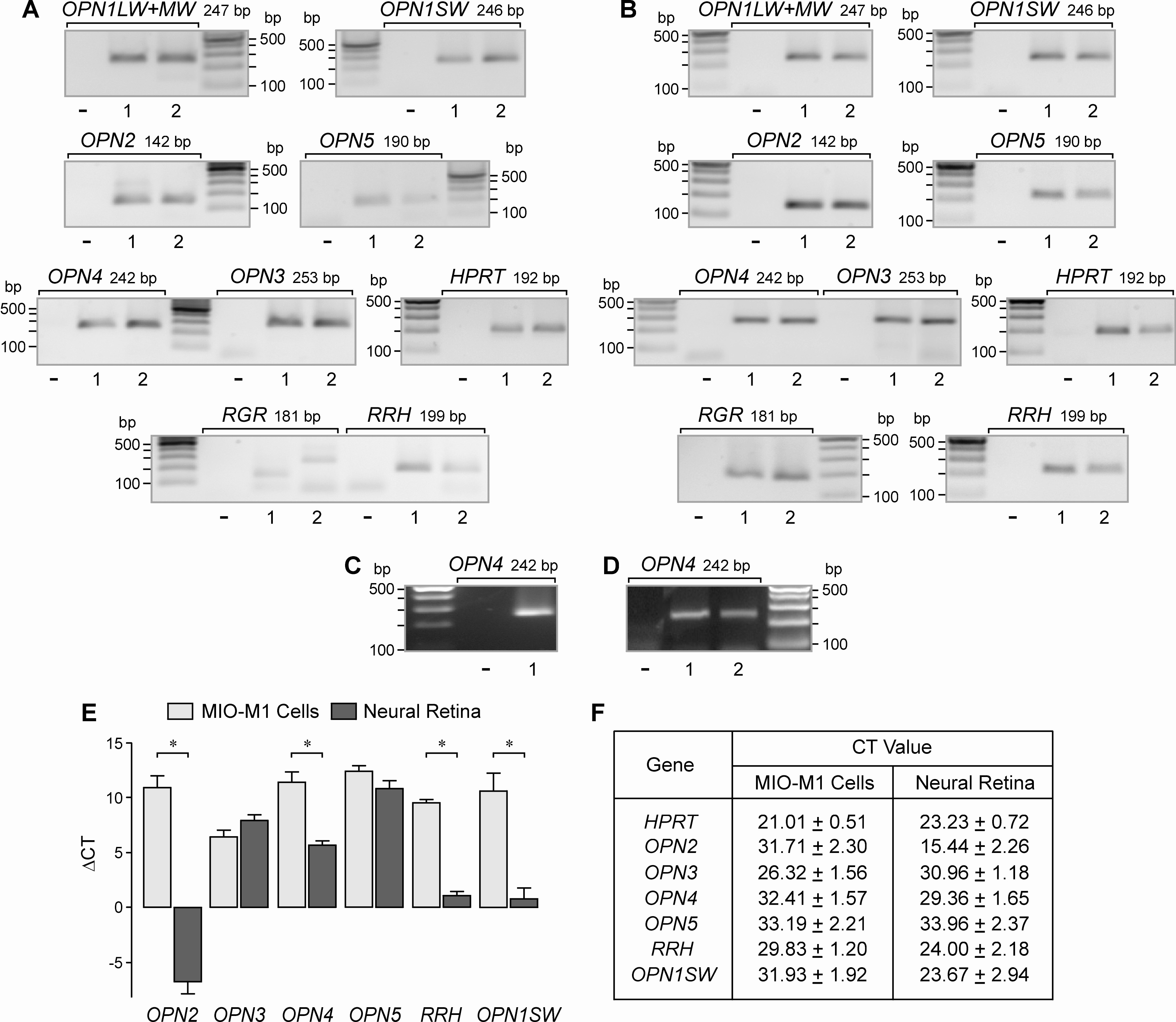
 Figure 3
of Hollborn, Mol Vis 2011; 17:2738-2750.
Figure 3
of Hollborn, Mol Vis 2011; 17:2738-2750.