Figure 2. Phenotype associated with retinal dehydrogenase 12 (RDH12) retinopathy. A: Fundus appearance in adults and older children shows dense intraretinal pigment migration, severe retinal pigment epithelium
atrophy, and arteriolar attenuation, with a severe atrophic pigmentary maculopathy (family 12, age 20 years). B: Para-arteriolar sparing of the intraretinal pigmentation was evident in six of 32 patients (white arrows, family 22, age
17.5 years). C: In children, retinal pigment epithelium atrophy with macular atrophy and minimal intraretinal pigmentation predominated
(family 17, age 8.5 years). D: Macular atrophy was often associated with striking yellow deposits (family 3, age 27 years). E: No detectable macular autofluorescence was visible on fundus autofluorescence imaging, corresponding to the severe macular
atrophy (family 11, age 11). F: Spectral domain optical coherence tomography demonstrated severe macular thinning, excavation, and distortion of the laminar
architecture (white arrow, family 22, age 17.5 years).
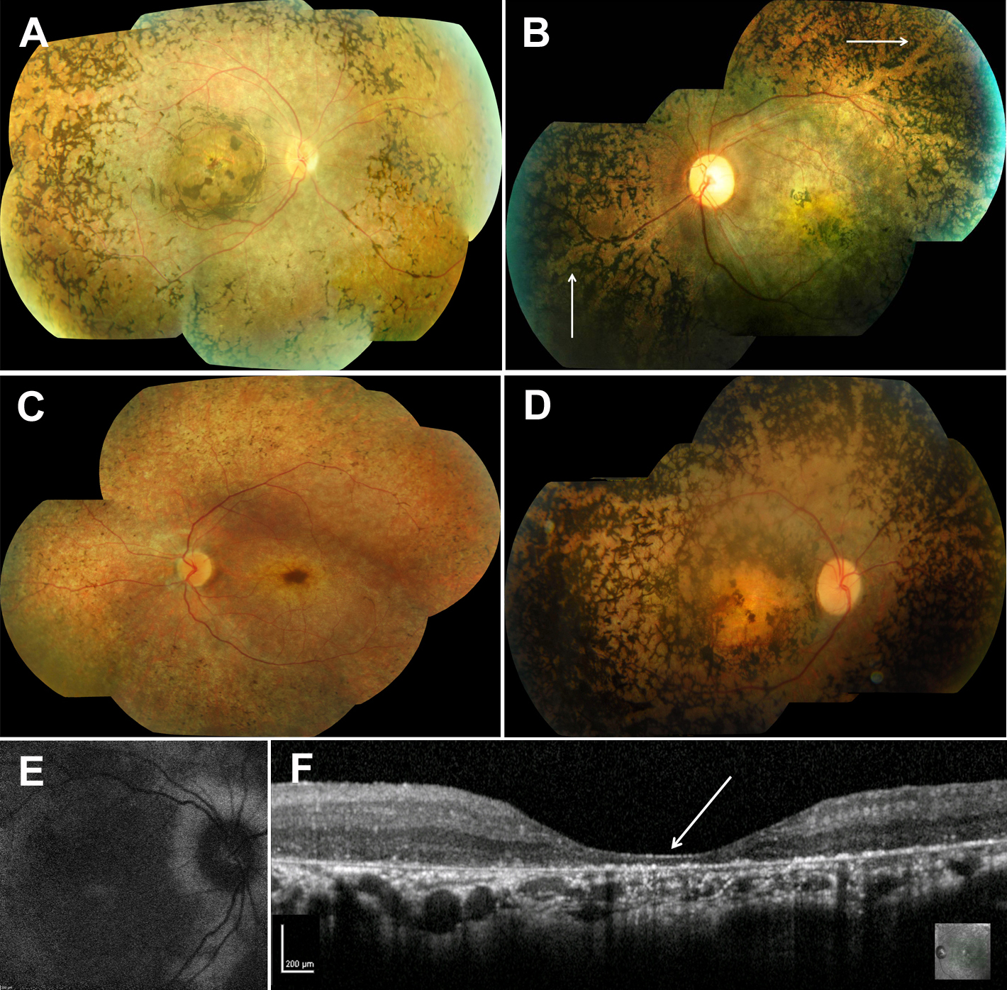
 Figure 2 of
Mackay, Mol Vis 2011; 17:2706-2716.
Figure 2 of
Mackay, Mol Vis 2011; 17:2706-2716.