Figure 4. Co-immunoprecipitation of
Pax6 and Pax6-variant. A: Rabbit anti-Gfp-Pax6 antibody
was used to do the IP experiment, and rabbit anti-Gfp-Pax6
antibody was used to do the western blotting. The inputs from
left to right are negative control, 6-day embryonic tissue
extract, Co-IP, and Pax6-his protein. Pax6 protein is obvious in
the 6-day embryonic tissue extract and the Co-IP of the input,
and lower than the heavy chain of the antibody. When using the
rabbit anti-Gfp-Pax6-varian antibody to do the western blotting,
the input from left to right is negative control, 6-day
embryonic tissue extract, Co-IP, and Pax6-varian-his protein.
Pax6-varian can be found in the 6-day embryonic tissue extract
and the Co-IP of the input. B: The anti-Gfp-Pax6-varian
antibody was used to do the IP experiment. When using
anti-Gfp-Pax6-varian antibody and corresponding input, Pax6
variant was probed in the 6-day embryonic tissue extract and the
Co-IP of the input; also, Pax6 was probed with an anti-Gfp-Pax6
antibody in the 6-day embryonic tissue extract and the Co-IP of
the input. The results show that both Pax6 and Pax6 variant
protein can be found in embryonic tissues of the Bufo raddei
Strauch, and a complex of these proteins was found in the Co-IP
of the input. WB: Western Blotting; WT: Wild Type.
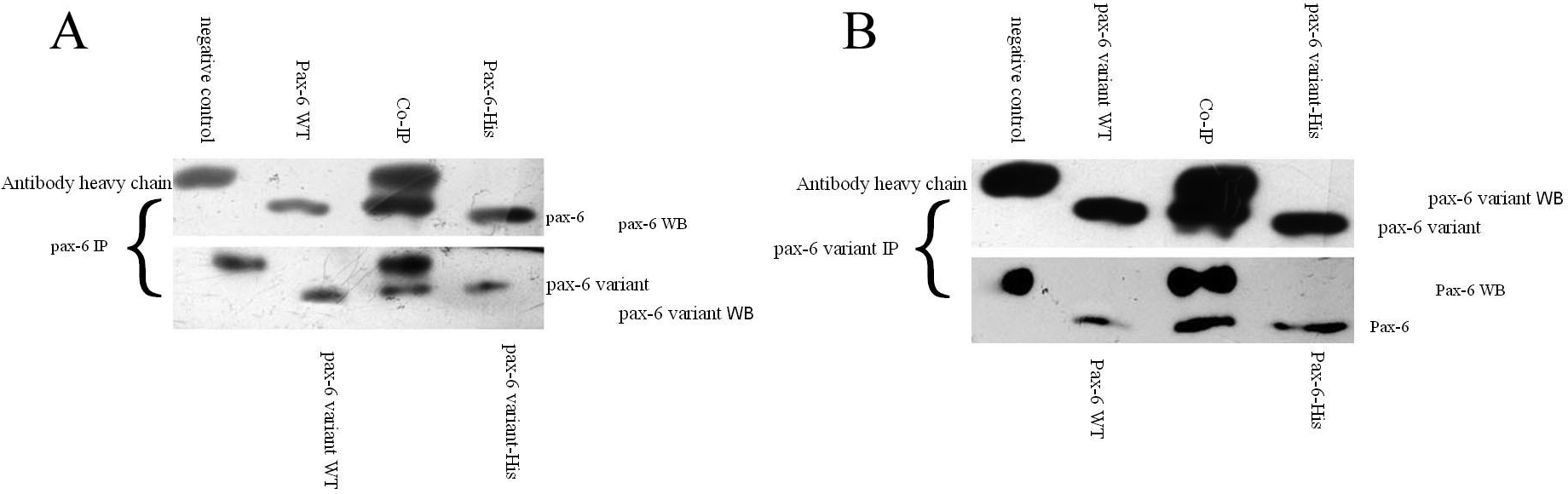
 Figure 4
of Ju, Mol Vis 2011; 17:2698-2705.
Figure 4
of Ju, Mol Vis 2011; 17:2698-2705.