Figure 1. Identification of
keratocytes. Inverted phase-contrast images (A) and
immune florescent vimentin staining (B) of human
keratocytes. Keratocytes were observed growing parallel to the
etched lines. Immunofluorescence with anti-vimentin antibody, a
general marker of keratocyte, showed the morphology. The nuclei
were stained with Hoechst 33342 (blue). Magnification, 200×.
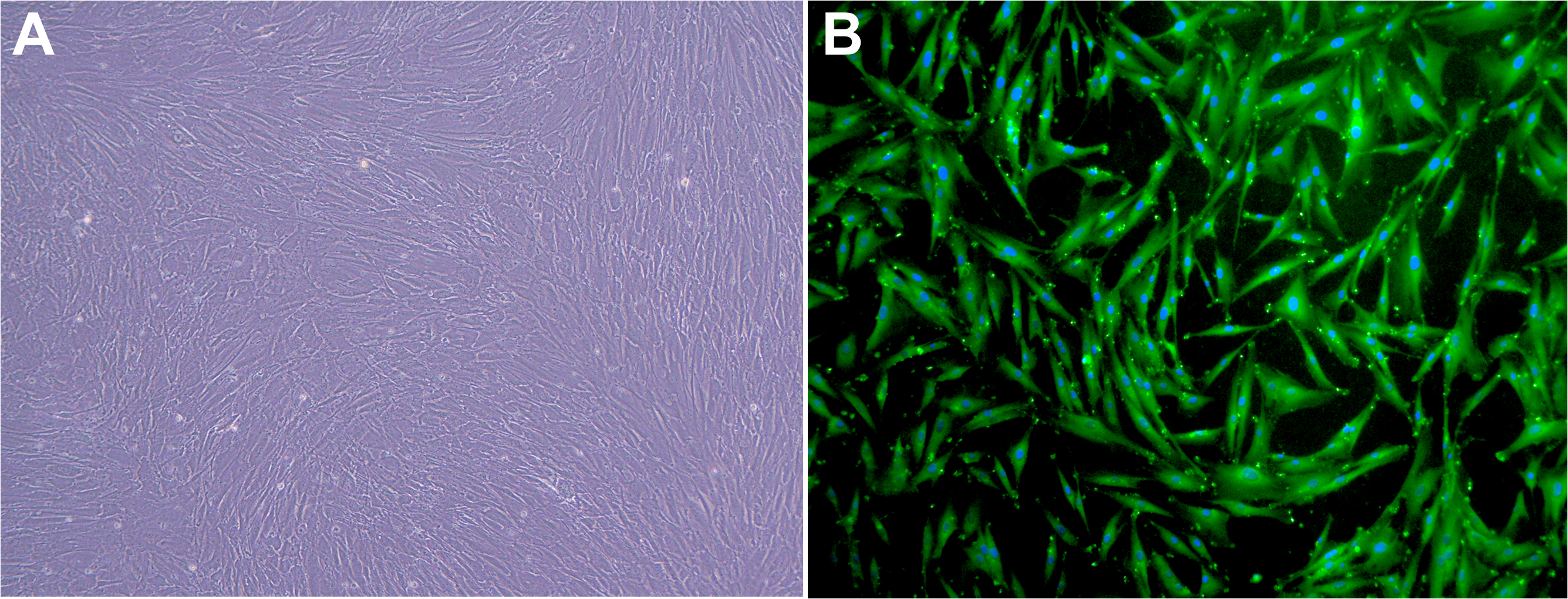
 Figure 1
of Yi, Mol Vis 2011; 17:2665-2671.
Figure 1
of Yi, Mol Vis 2011; 17:2665-2671.