Figure 1. Confocal images of Ctns-\- mouse cornea. Confocal images of the same Ctns−/− cornea over time at 3 months (A), 6 months (B), 8 months (C), and 10 months of age (D). Each panel shows a xy (upper) slice through a 3D stack. Cystine crystals were identified as small, 20 µm long, needle-like
crystals in the peripheral and central cornea. Note that cystine crystals increase progressively in quantity up to 8 months
of age (A, B, and C), but at 10 months, the cornea scarred and showed increased opacity.
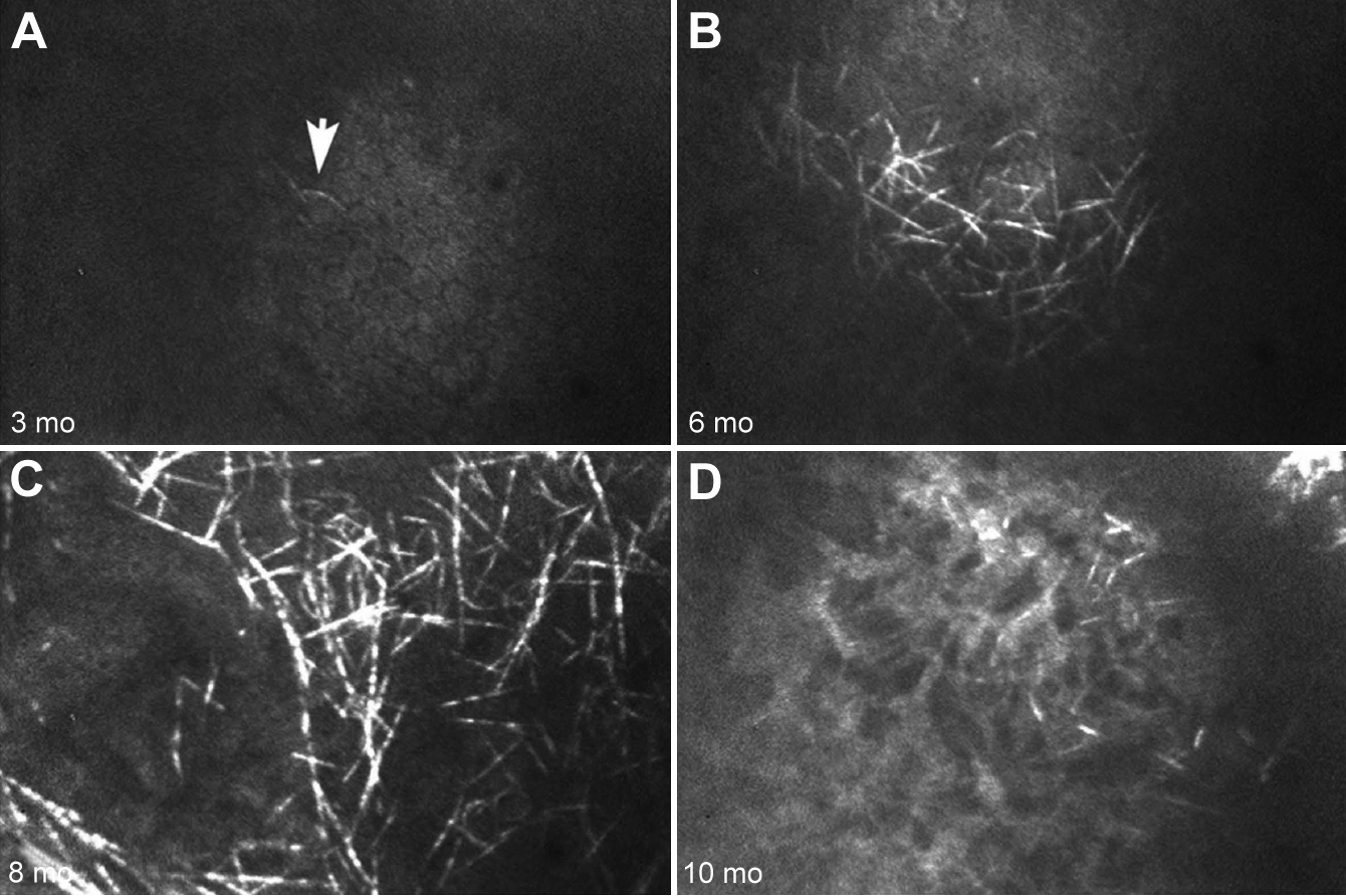
 Figure 1 of
Simpson, Mol Vis 2011; 17:2649-2654.
Figure 1 of
Simpson, Mol Vis 2011; 17:2649-2654.