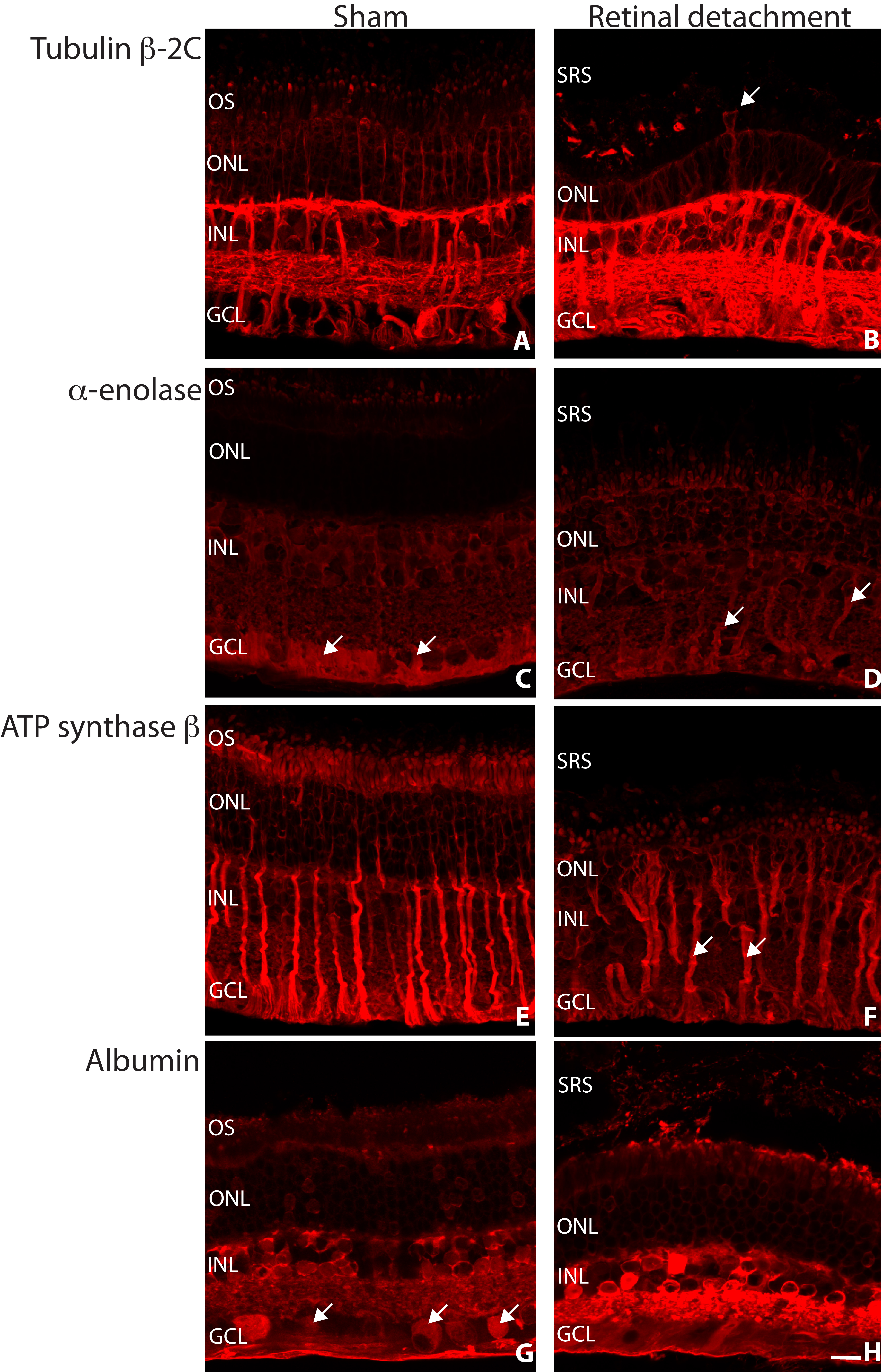
Figure 5. Laser scanning confocal
images of sham (A, C, E, G) and
detached (B, D, F, H) rabbit
retina labeled with antibodies for tubulin β-2C (A, B),
α-enolase (C, D), ATP synthase β (E, F),
and albumin (G, H) all in red. Müller cells are
often observed extending into the subretinal space following
retinal detachment (B, arrow). Labeling for α-enolase was
concentrated in the Müller cell endfeet region in sham retina,
which is possibly redistributed within this cell type following
retinal detachment (C, D, arrows). The Müller
cells remained relatively brightly labeled for ATP synthase
subunit β following retinal detachment (F, arrows).
Variable albumin labeling of the ganglion cells was observed in
sham retina (G, arrows). The albumin labeling that
occurred intracellularly was shown to increase in intensity
following retinal detachment (G, H).
Abbreviations: OS represents outer segments; ONL represents
outer nuclear layer; INL represents inner nuclear layer; GCL
represents ganglion cell layer; SRS represents subretinal space.
Scale bar 20 µm.

 Figure 5
of Mandal, Mol Vis 2011; 17:2634-2648.
  Figure 5
of Mandal, Mol Vis 2011; 17:2634-2648.