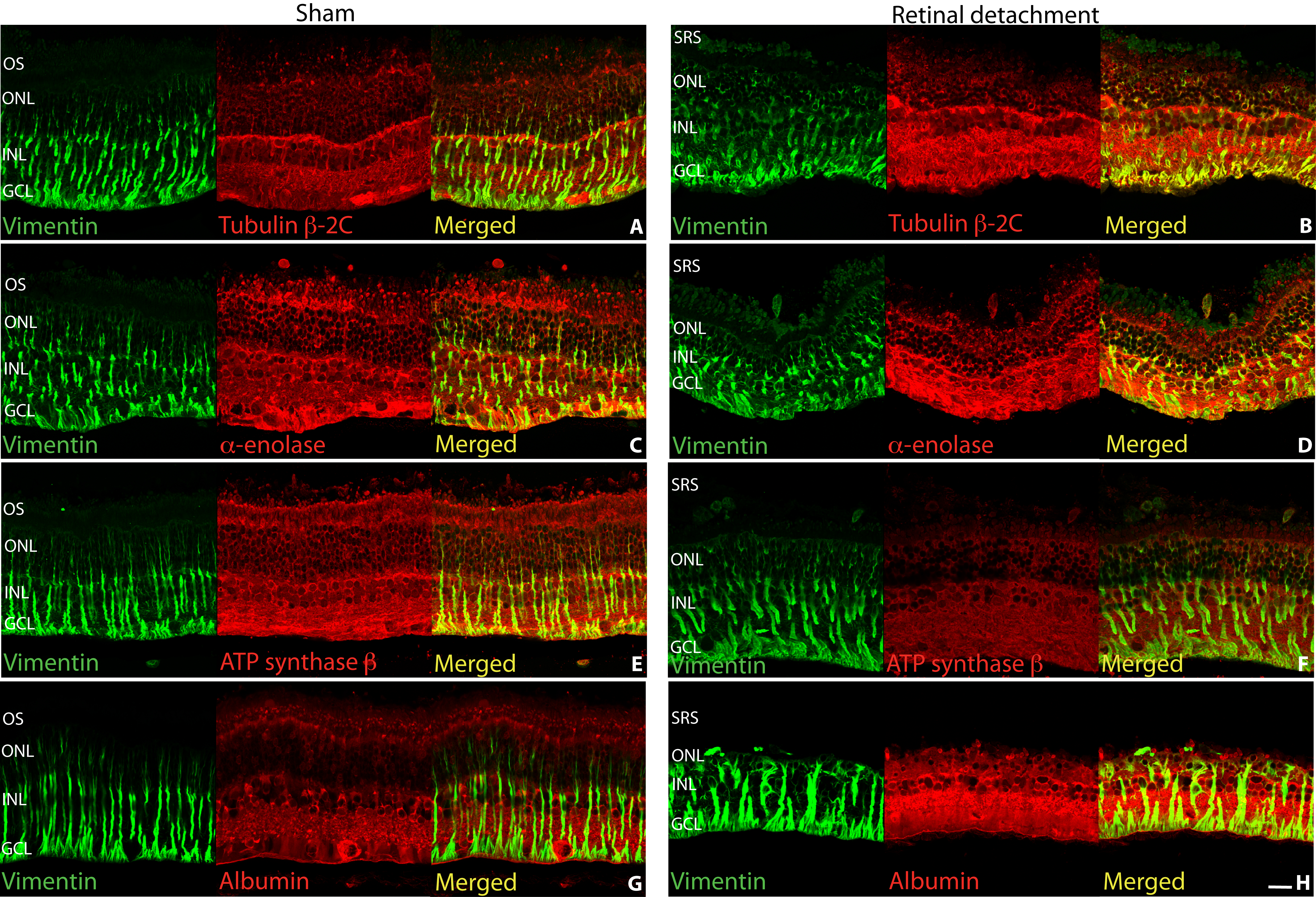
Figure 4. Laser scanning confocal
images of sham (A, C, E, G) and
detached (B, D, F, H) rabbit
retina labeled with antibodies for vimentin (A-H,
green), tubulin β-2C (A, B), α-enolase (C,
D), ATP synthase (E, F), and albumin (G,
H) all in red. Colocalization of the respective proteins
in the Müller cells is suggested by greenish yellow to yellow
color. Abbreviations: OS represents outer segments; ONL
represents outer nuclear layer; INL represents inner nuclear
layer; GCL represents ganglion cell layer; SRS represents
subretinal space. Scale bar 20 µm.

 Figure 4
of Mandal, Mol Vis 2011; 17:2634-2648.
  Figure 4
of Mandal, Mol Vis 2011; 17:2634-2648.