Figure 3. Label free imaging of the
trabecular meshwork of a human cadaver eye using two photon
autofluorescence (TPAF) and Coherent Anti-Stokes Raman
Scattering (CARS) microscopy. (Green: TPAF, Red: CARS) The
arrows indicate the TM cells that reside in the interstitial
areas within the collagen extracellular matrix (EM) structure.
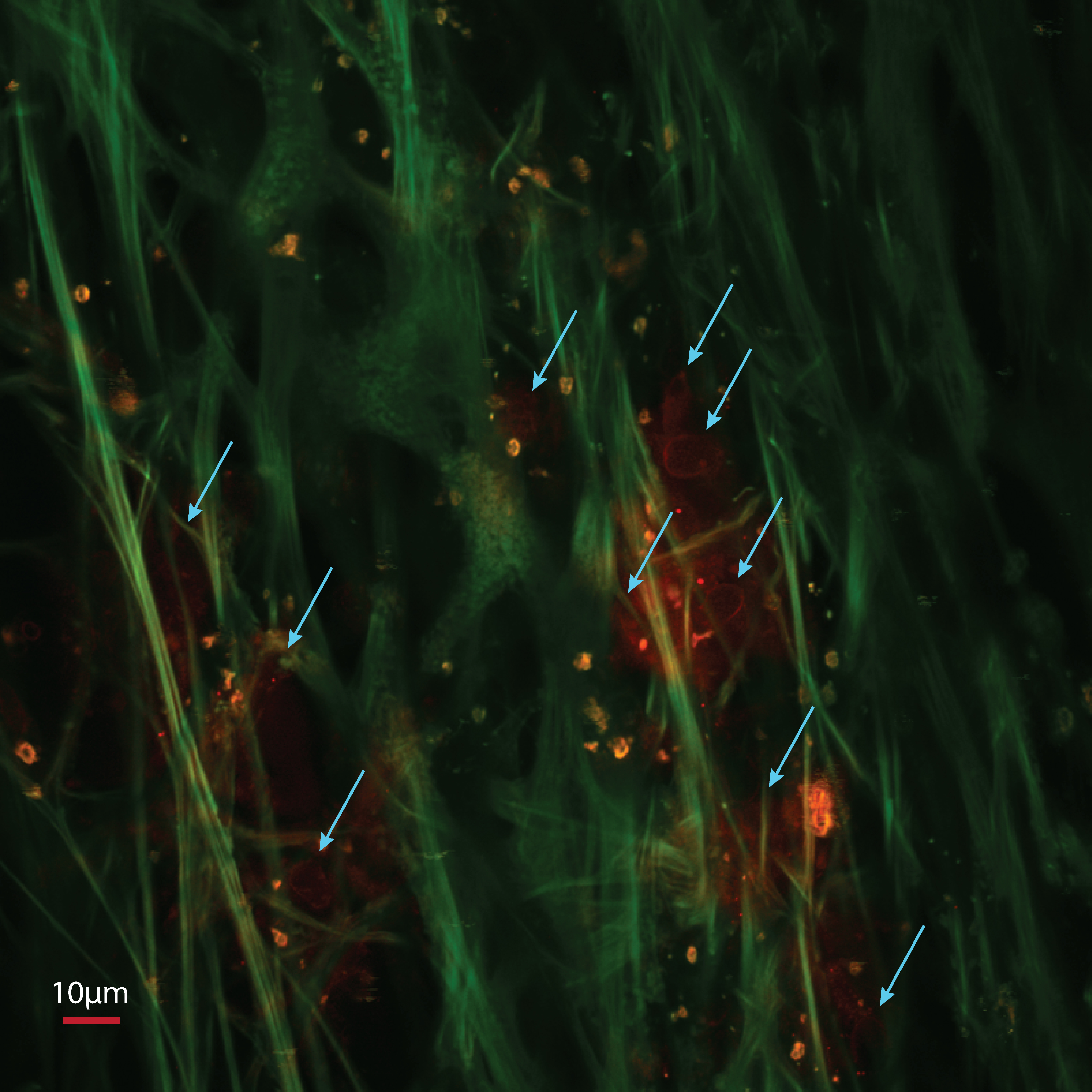
 Figure 3
of Lei, Mol Vis 2011; 17:2628-2633.
Figure 3
of Lei, Mol Vis 2011; 17:2628-2633.