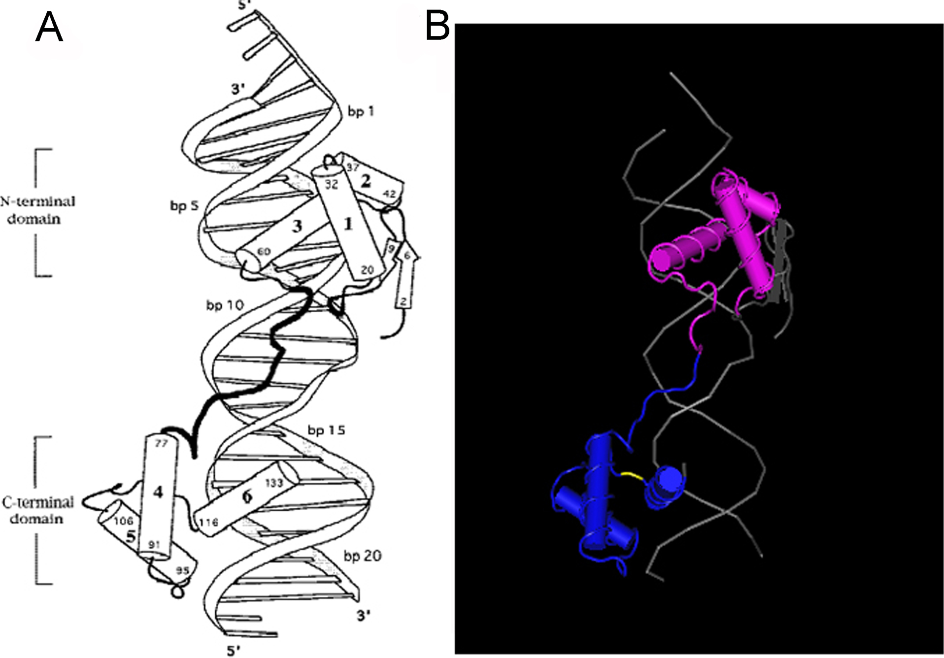
Figure 5. The paired domain of PAX6.
A: Sketch of the PAX6 paired domain–DNA complex.
Cylinders represent α helices; arrows represent β strands.
Helices 1–6 are labeled; residue numbers indicate termini of the
corresponding secondary structure elements.
B:
Cn3D
display for the paired domain of PAX6. The yellow segment
represents the mutation region (COOH-terminal region in PAX6
paired domain).
 Figure 5
of Yan, Mol Vis 2011; 17:2612-2617.
Figure 5
of Yan, Mol Vis 2011; 17:2612-2617.