Figure 1. Corneal fluorescein
staining photographs. Slit-lamp microscopic and fluorescein
staining photographs from a representative patient with dry eye
related to GVHD (A) and a control (B). Green
punctate positive fluorescein staining in A highlights
the erosion of the corneal epithelium; B shows an intact
corneal epithelium in a control.
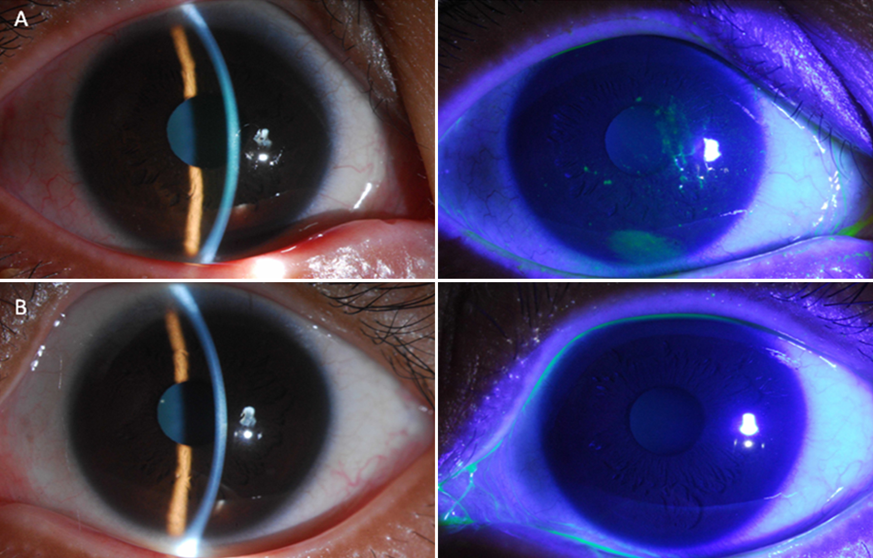
 Figure 1
of He, Mol Vis 2011; 17:2605-2611.
Figure 1
of He, Mol Vis 2011; 17:2605-2611.