Figure 7. The immunoreactivity of transplanted ciliary epithelium (CE)-derived cells in the neuroretina. A: Microphotograph (13.3 µm confocal stack) showing red CM-DiI-labeled cells positive for recoverin (green, arrows) in the
outer nuclear layer (ONL). B: Recoverin-only labeling of the same confocal stack as in A. The arrows point to the transplanted cells. C: A 0.7 µm confocal slice from the boxed area in A, showing double-labeled CM-DiI/recoverin positive cells (arrows) D: Green recoverin labeling in the same area (arrow). E: Red CM-DiI labeling and 4',6-diamidino-2-phenylindole (DAPI) nuclear staining of the same area. A, F, G: CM-DiI-labeled, protein kinase α (PKCα)-positive cell (green) in the inner plexiform layer (arrows). A, H, I: HuC/D (green) and CM-DiI-positive cell in the ganglion cell layer (GCL; arrow in H and I). J, K: Calbindin (green) and CM-DiI-labeled cells in the GCL (arrows). The inset is at a higher magnification with visible processes
(arrowhead). The nuclei are labeled with DAPI (blue). Inner nuclear layer (INL).
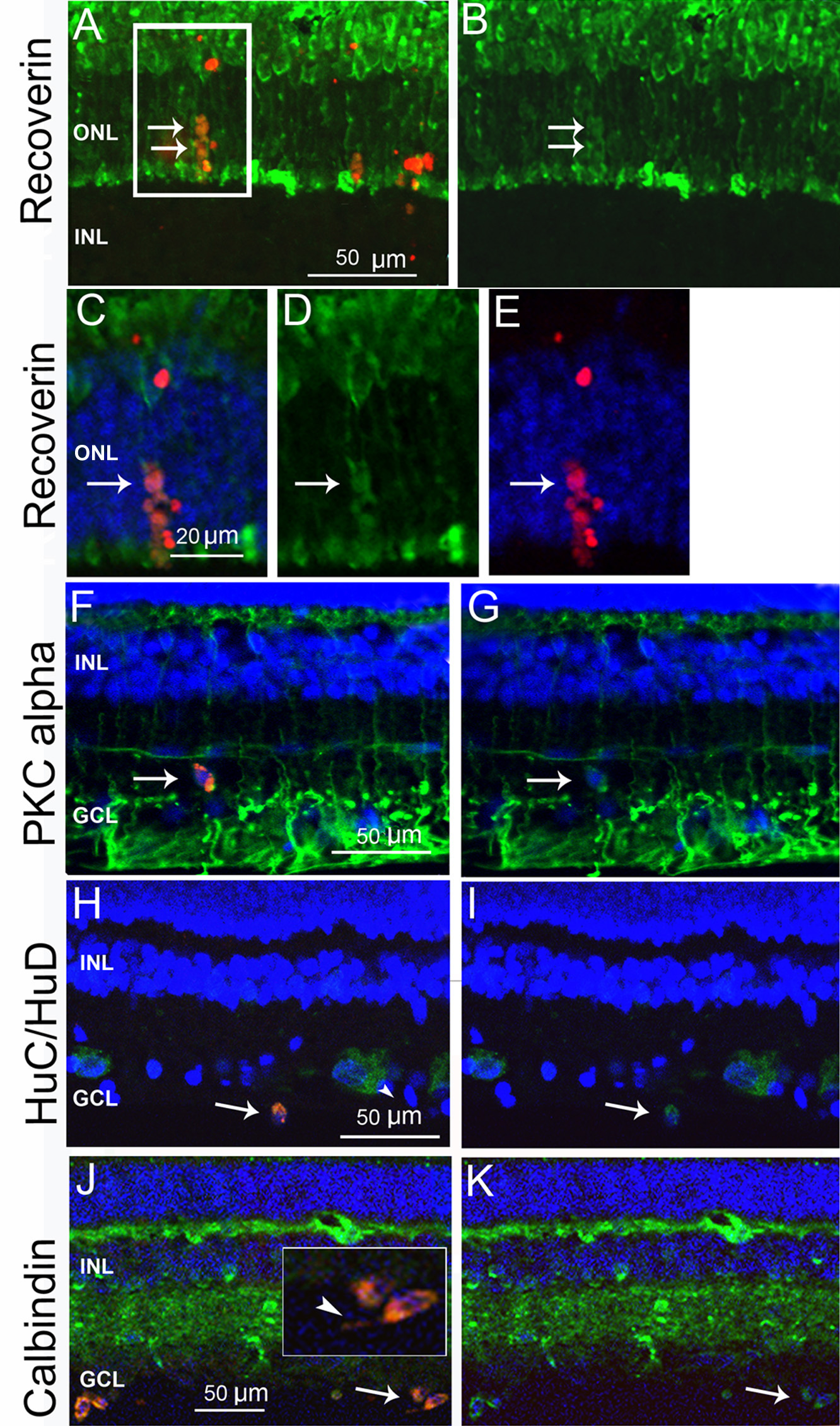
 Figure 7 of
Guduric-Fuchs, Mol Vis 2011; 17:2580-2595.
Figure 7 of
Guduric-Fuchs, Mol Vis 2011; 17:2580-2595.