Figure 6. Analysis of migration, proliferation, and death of porcine ciliary epithelium (CE)-derived cells after subretinal transplantation.
A: Quantification of the transplanted cells that had migrated into the neuroretina. CM-DiI-labeled cells were counted in 20
random sections from each eye. The middle third, containing the optic nerve, was considered to be the central retina and the
two peripheral thirds, including the ora serrata, were considered to be the peripheral retina. The results are presented as
the mean±SEM B, C: Cell proliferation assessed by Ki67 labeling in transplanted retinas. CM-DiI-positive cell aggregates in the subretinal
space (red in B) contained rare Ki67-labeled cells (green, arrow in C), eight days after transplantation. D, E: Phagocytosis of transplanted cells by macrophages. CM-DiI-labeled particles (red in D, arrow) contained within isolectin B4-positive macrophages (green in E, arrow). The nuclei are labeled with 4',6-diamidino-2-phenylindole (DAPI; blue). Outer nuclear layer (ONL); inner nuclear
layer (INL); and subretinal space (SS).
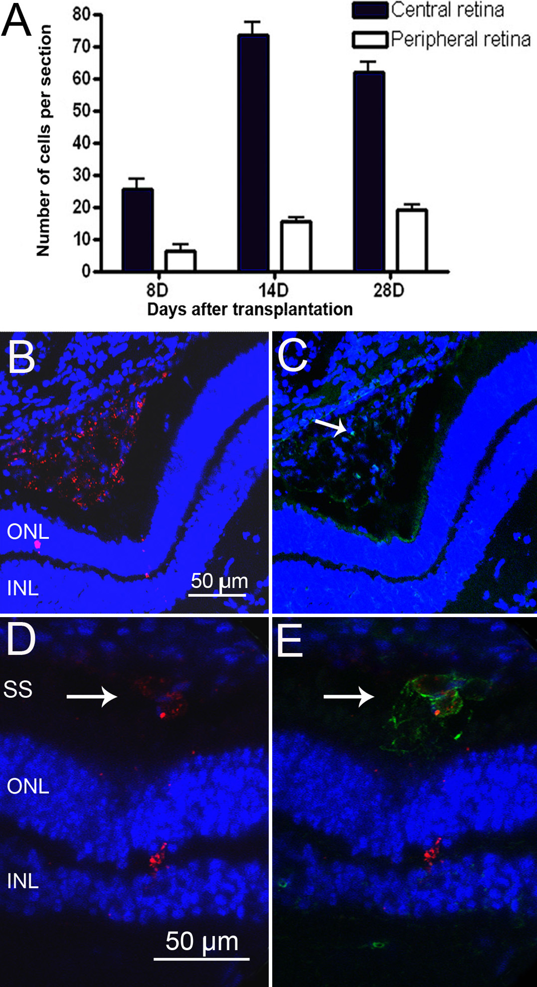
 Figure 6 of
Guduric-Fuchs, Mol Vis 2011; 17:2580-2595.
Figure 6 of
Guduric-Fuchs, Mol Vis 2011; 17:2580-2595.