Figure 5. Microphotographs of red
CM-DiI-labeled ciliary epithelium (CE)-derived cells in the
retinal pigment epithelium (RPE) layer. A: Red-labeled
pigmented CE-derived cells localized to the RPE layer, which
were negative for RPE65 (arrows). B: At the same time
point, transplanted red-labeled RPE65-positive cells were also
found (red and green merged in B and green RPE65
labeling only in C, arrows). D-F: Two
weeks following transplantation, CM-DiI-labeled cells in the RPE
layer were strongly (arrow) and weakly (arrowhead) positive for
RPE65. G-I: Four weeks after transplantation, the
RPE appeared uneven and multilayered (arrows). Nuclei are
labeled with 4',6-diamidino-2-phenylindole (DAPI; blue).
Bright-field images are merged with the dark field in A,
G, H, and I. Subretinal space (SS); and
outer nuclear layer (ONL).
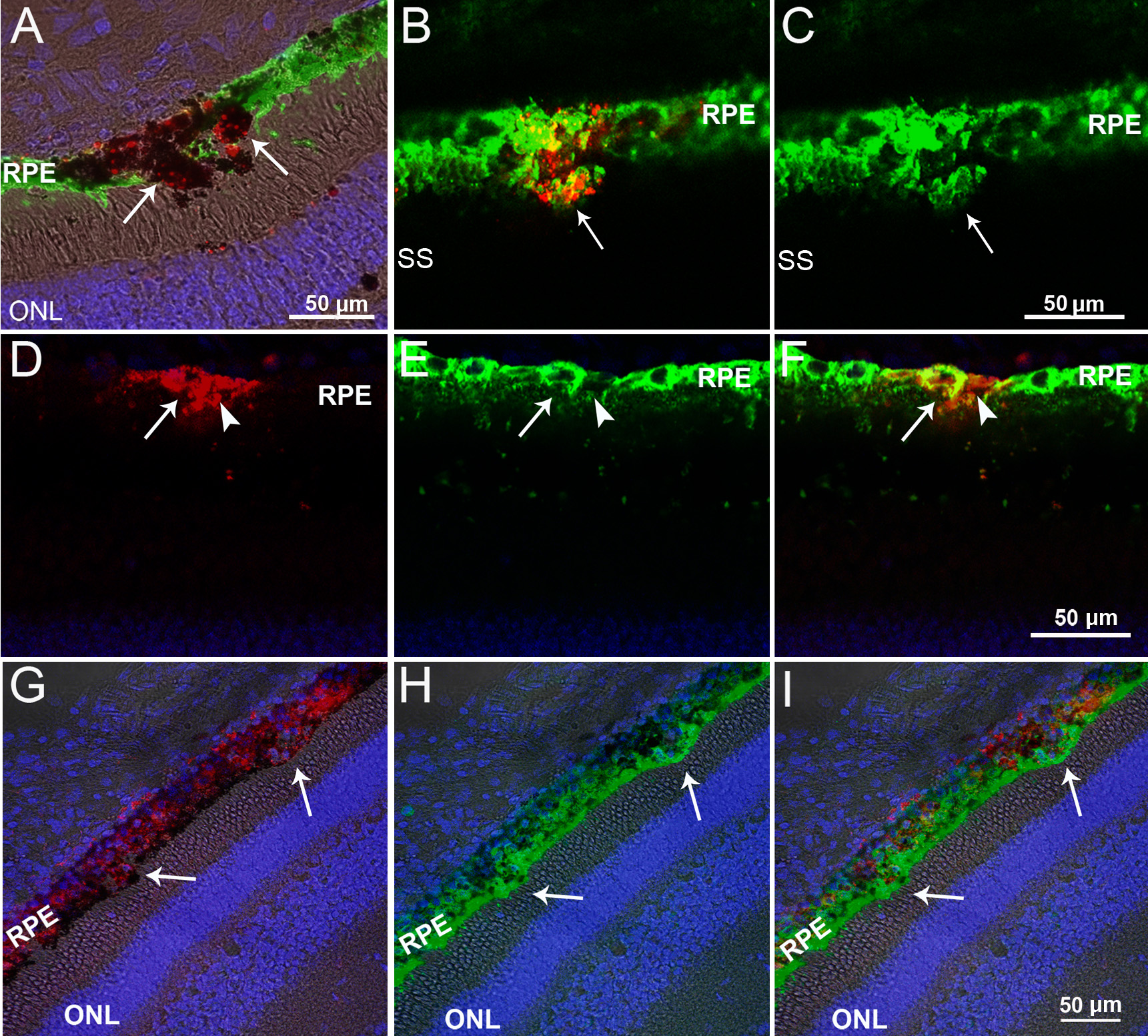
 Figure 5
of Guduric-Fuchs, Mol Vis 2011;
17:2580-2595.
Figure 5
of Guduric-Fuchs, Mol Vis 2011;
17:2580-2595.