Figure 4. CM-DiI labeling of ciliary epithelium (CE)-derived cells. Dissociated cells at passage 1 were labeled and cultured in a suspension
culture to form spheres (A), or were plated on poly-D-Lysine, laminin coated coverslips in differentiation conditions. The CM-DiI label was retained
for up to 10 days in proliferating (spheres depicted by arrows in A), and up to four weeks in differentiating conditions (arrows in B) in vitro. C, D: Microphotographs of grafted CM-DiI-labeled CE-derived cells (red) in the recipient retina 10 min after subretinal injection.
C: CM-DiI labeling merged with nuclear DAPI staining. D: 4',6-diamidino-2-phenylindole (DAPI) staining only. Red CM-DiI-labeled cells were found between the RPE (arrow) and the
ONL. Retinal pigment epithelium (RPE); outer nuclear layer (ONL).
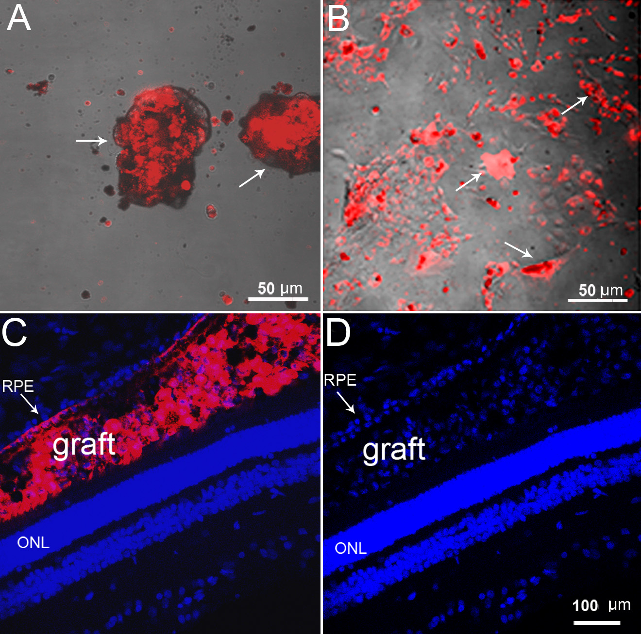
 Figure 4 of
Guduric-Fuchs, Mol Vis 2011; 17:2580-2595.
Figure 4 of
Guduric-Fuchs, Mol Vis 2011; 17:2580-2595.